Our official English website, www.x-mol.net, welcomes your
feedback! (Note: you will need to create a separate account there.)
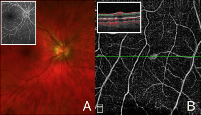
Use of optical coherence tomography angiography in the diagnosis of small retina lesions in Von Hippel–Lindau disease
Eye ( IF 2.8 ) Pub Date : 2020-02-10 , DOI: 10.1038/s41433-020-0792-5 Lindsay Y Chun 1 , Nathalie Massamba 1 , Megan R Silas 1 , Michael P Blair 1 , Seenu M Hariprasad 1 , Dimitra Skondra 1, 2
Eye ( IF 2.8 ) Pub Date : 2020-02-10 , DOI: 10.1038/s41433-020-0792-5 Lindsay Y Chun 1 , Nathalie Massamba 1 , Megan R Silas 1 , Michael P Blair 1 , Seenu M Hariprasad 1 , Dimitra Skondra 1, 2
Affiliation
|
Von Hippel–Lindau disease (VHL) is a neoplastic disorder that can lead to the multisystem formation of tumors and cysts in the central nervous system, adrenal glands, kidneys, pancreas, reproductive system, and retina [1]. The earliest ocular findings of VHL are retinal capillary hemangiomas, and loss of vision can result from tumor growth, extravasation of fluid and lipid exudates from the lesions, and retinal detachment. The evaluation of retinal manifestations of VHL includes the clinical exam and fundus fluorescein angiography (FFA), but FFA is invasive and findings of small lesions can be equivocal, making the decision for treatment challenging. Optical coherence tomography with angiography (OCTA) is a novel, noninvasive imaging technique that can provide in vivo images of the retinal vasculature with higher resolution than conventional techniques. However, the ability of OCTA to confirm both the presence and absence of potential retinal lesions seen with FFA in VHL has not yet been described. We have observed two cases of retinal hemangiomas with a systemic history of VHL.
中文翻译:

光学相干断层扫描血管造影术在 Von Hippel-Lindau 病视网膜小病变诊断中的应用
Von Hippel-Lindau 病 (VHL) 是一种肿瘤性疾病,可导致中枢神经系统、肾上腺、肾脏、胰腺、生殖系统和视网膜中肿瘤和囊肿的多系统形成[1]。VHL 最早的眼部表现是视网膜毛细血管瘤,视力丧失可由肿瘤生长、病变处的液体和脂质渗出物外渗以及视网膜脱离引起。VHL 视网膜表现的评估包括临床检查和眼底荧光素血管造影 (FFA),但 FFA 是侵入性的,小病变的发现可能不明确,这使得治疗决策具有挑战性。光学相干断层扫描与血管造影 (OCTA) 是一种新颖的、一种无创成像技术,可以提供比传统技术分辨率更高的视网膜脉管系统的活体图像。然而,尚未描述 OCTA 确认 VHL 中 FFA 观察到的潜在视网膜病变存在和不存在的能力。我们观察到两例具有 VHL 全身病史的视网膜血管瘤。
更新日期:2020-02-10
中文翻译:
光学相干断层扫描血管造影术在 Von Hippel-Lindau 病视网膜小病变诊断中的应用
Von Hippel-Lindau 病 (VHL) 是一种肿瘤性疾病,可导致中枢神经系统、肾上腺、肾脏、胰腺、生殖系统和视网膜中肿瘤和囊肿的多系统形成[1]。VHL 最早的眼部表现是视网膜毛细血管瘤,视力丧失可由肿瘤生长、病变处的液体和脂质渗出物外渗以及视网膜脱离引起。VHL 视网膜表现的评估包括临床检查和眼底荧光素血管造影 (FFA),但 FFA 是侵入性的,小病变的发现可能不明确,这使得治疗决策具有挑战性。光学相干断层扫描与血管造影 (OCTA) 是一种新颖的、一种无创成像技术,可以提供比传统技术分辨率更高的视网膜脉管系统的活体图像。然而,尚未描述 OCTA 确认 VHL 中 FFA 观察到的潜在视网膜病变存在和不存在的能力。我们观察到两例具有 VHL 全身病史的视网膜血管瘤。


 京公网安备 11010802027423号