当前位置:
X-MOL 学术
›
NMR Biomed.
›
论文详情
Our official English website, www.x-mol.net, welcomes your
feedback! (Note: you will need to create a separate account there.)
Resolution-dependent influences of compressed sensing in quantitative T2 mapping of articular cartilage.
NMR in Biomedicine ( IF 2.7 ) Pub Date : 2020-02-10 , DOI: 10.1002/nbm.4260 Nian Wang 1, 2 , Farid Badar 3 , Yang Xia 3
NMR in Biomedicine ( IF 2.7 ) Pub Date : 2020-02-10 , DOI: 10.1002/nbm.4260 Nian Wang 1, 2 , Farid Badar 3 , Yang Xia 3
Affiliation
|
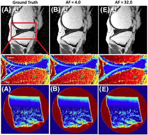
This study evaluates the resolution‐dependent influences of compressed sensing (CS) in MRI quantification of T2 mapping in articular cartilage with osteoarthritis (OA). T2‐weighed 2D experiments of healthy and OA cartilage were fully sampled in k‐space with five echo times at both 17.6 μm and 195.3 μm in‐plane resolutions; termed as microscopic MRI (μMRI) and macroscopic MRI (mMRI) respectively. These fully sampled k‐space data were under‐sampled at various 2D CS accelerating factors (AF = 4–32). The under‐sampled data were reconstructed individually into 2D images using nonlinear reconstruction, which were used to calculate the T2 maps. The bulk and zonal variations of T2 values in cartilage were evaluated at different AFs. The study finds that the T2 images at AFs up to 8 preserved major visual information and produced negligible artifacts for μMRI. The T2 values remained accurate for different sub‐tissue zones at various AFs. The absolute difference between the CS (AF up to 32) and the Ground Truth (i.e., using 100% of the k‐space data) of the mean T2 values through the whole tissue depth was higher in mMRI versus μMRI. For mMRI (where the resolution mimics the clinical MRI of human cartilage), the quantitative T2 mapping at AFs up to 4 showed negligible variations. This study demonstrates that both clinical MRI and μMRI can benefit from the use of CS in image acquisition, and μMRI benefits more from the use of CS by acquiring much less data, without losing significant accuracy in the quantification of T2 maps in osteoarthritic cartilage.
中文翻译:

压缩感知在关节软骨定量 T2 映射中的分辨率相关影响。
本研究评估了压缩感知 (CS) 在骨关节炎 (OA) 关节软骨 T2 映射的 MRI 量化中的分辨率依赖性影响。健康和 OA 软骨的 T2 加权 2D 实验在 k 空间中以 17.6 μm 和 195.3 μm 面内分辨率的五个回波时间完全采样;分别称为显微MRI(μMRI)和宏观MRI(mMRI)。这些完全采样的 k 空间数据在各种 2D CS 加速因子(AF = 4-32)下采样不足。使用非线性重建将欠采样数据单独重建为二维图像,用于计算 T2 图。在不同的 AF 下评估了软骨中 T2 值的整体和带状变化。该研究发现,AFs 的 T2 图像最多 8 个保留了主要的视觉信息,并且对 μMRI 产生的伪影可以忽略不计。对于不同 AF 的不同亚组织区域,T2 值保持准确。与 μMRI 相比,mMRI 中整个组织深度的平均 T2 值的 CS(AF 高达 32)和 Ground Truth(即使用 100% 的 k 空间数据)之间的绝对差异更高。对于 mMRI(其分辨率模拟人类软骨的临床 MRI),AFs 的定量 T2 映射高达 4 显示出可以忽略不计的变化。这项研究表明,临床 MRI 和 μMRI 都可以从在图像采集中使用 CS 中受益,并且 μMRI 通过获取更少的数据而从 CS 的使用中受益更多,而不会失去骨关节炎软骨中 T2 图量化的显着准确性。对于不同 AF 的不同亚组织区域,T2 值保持准确。与 μMRI 相比,mMRI 中整个组织深度的平均 T2 值的 CS(AF 高达 32)和 Ground Truth(即使用 100% 的 k 空间数据)之间的绝对差异更高。对于 mMRI(其分辨率模拟人类软骨的临床 MRI),AFs 的定量 T2 映射高达 4 显示出可以忽略不计的变化。这项研究表明,临床 MRI 和 μMRI 都可以从在图像采集中使用 CS 中受益,并且 μMRI 通过获取更少的数据而从 CS 的使用中受益更多,而不会失去骨关节炎软骨中 T2 图量化的显着准确性。对于不同 AF 的不同亚组织区域,T2 值保持准确。与 μMRI 相比,mMRI 中整个组织深度的平均 T2 值的 CS(AF 高达 32)和 Ground Truth(即使用 100% 的 k 空间数据)之间的绝对差异更高。对于 mMRI(其分辨率模拟人类软骨的临床 MRI),AFs 的定量 T2 映射高达 4 显示出可以忽略不计的变化。这项研究表明,临床 MRI 和 μMRI 都可以从在图像采集中使用 CS 中受益,并且 μMRI 通过获取更少的数据而从 CS 的使用中受益更多,而不会失去骨关节炎软骨中 T2 图量化的显着准确性。使用 100% 的 k 空间数据)整个组织深度的平均 T2 值在 mMRI 中高于 μMRI。对于 mMRI(其分辨率模拟人类软骨的临床 MRI),AFs 的定量 T2 映射高达 4 显示出可以忽略不计的变化。这项研究表明,临床 MRI 和 μMRI 都可以从在图像采集中使用 CS 中受益,并且 μMRI 通过获取更少的数据而从 CS 的使用中受益更多,而不会失去骨关节炎软骨中 T2 图量化的显着准确性。使用 100% 的 k 空间数据)整个组织深度的平均 T2 值在 mMRI 中高于 μMRI。对于 mMRI(其分辨率模拟人类软骨的临床 MRI),AFs 的定量 T2 映射高达 4 显示出可以忽略不计的变化。这项研究表明,临床 MRI 和 μMRI 都可以从在图像采集中使用 CS 中受益,并且 μMRI 通过获取更少的数据而从 CS 的使用中受益更多,而不会失去骨关节炎软骨中 T2 图量化的显着准确性。
更新日期:2020-02-10
中文翻译:
压缩感知在关节软骨定量 T2 映射中的分辨率相关影响。
本研究评估了压缩感知 (CS) 在骨关节炎 (OA) 关节软骨 T2 映射的 MRI 量化中的分辨率依赖性影响。健康和 OA 软骨的 T2 加权 2D 实验在 k 空间中以 17.6 μm 和 195.3 μm 面内分辨率的五个回波时间完全采样;分别称为显微MRI(μMRI)和宏观MRI(mMRI)。这些完全采样的 k 空间数据在各种 2D CS 加速因子(AF = 4-32)下采样不足。使用非线性重建将欠采样数据单独重建为二维图像,用于计算 T2 图。在不同的 AF 下评估了软骨中 T2 值的整体和带状变化。该研究发现,AFs 的 T2 图像最多 8 个保留了主要的视觉信息,并且对 μMRI 产生的伪影可以忽略不计。对于不同 AF 的不同亚组织区域,T2 值保持准确。与 μMRI 相比,mMRI 中整个组织深度的平均 T2 值的 CS(AF 高达 32)和 Ground Truth(即使用 100% 的 k 空间数据)之间的绝对差异更高。对于 mMRI(其分辨率模拟人类软骨的临床 MRI),AFs 的定量 T2 映射高达 4 显示出可以忽略不计的变化。这项研究表明,临床 MRI 和 μMRI 都可以从在图像采集中使用 CS 中受益,并且 μMRI 通过获取更少的数据而从 CS 的使用中受益更多,而不会失去骨关节炎软骨中 T2 图量化的显着准确性。对于不同 AF 的不同亚组织区域,T2 值保持准确。与 μMRI 相比,mMRI 中整个组织深度的平均 T2 值的 CS(AF 高达 32)和 Ground Truth(即使用 100% 的 k 空间数据)之间的绝对差异更高。对于 mMRI(其分辨率模拟人类软骨的临床 MRI),AFs 的定量 T2 映射高达 4 显示出可以忽略不计的变化。这项研究表明,临床 MRI 和 μMRI 都可以从在图像采集中使用 CS 中受益,并且 μMRI 通过获取更少的数据而从 CS 的使用中受益更多,而不会失去骨关节炎软骨中 T2 图量化的显着准确性。对于不同 AF 的不同亚组织区域,T2 值保持准确。与 μMRI 相比,mMRI 中整个组织深度的平均 T2 值的 CS(AF 高达 32)和 Ground Truth(即使用 100% 的 k 空间数据)之间的绝对差异更高。对于 mMRI(其分辨率模拟人类软骨的临床 MRI),AFs 的定量 T2 映射高达 4 显示出可以忽略不计的变化。这项研究表明,临床 MRI 和 μMRI 都可以从在图像采集中使用 CS 中受益,并且 μMRI 通过获取更少的数据而从 CS 的使用中受益更多,而不会失去骨关节炎软骨中 T2 图量化的显着准确性。使用 100% 的 k 空间数据)整个组织深度的平均 T2 值在 mMRI 中高于 μMRI。对于 mMRI(其分辨率模拟人类软骨的临床 MRI),AFs 的定量 T2 映射高达 4 显示出可以忽略不计的变化。这项研究表明,临床 MRI 和 μMRI 都可以从在图像采集中使用 CS 中受益,并且 μMRI 通过获取更少的数据而从 CS 的使用中受益更多,而不会失去骨关节炎软骨中 T2 图量化的显着准确性。使用 100% 的 k 空间数据)整个组织深度的平均 T2 值在 mMRI 中高于 μMRI。对于 mMRI(其分辨率模拟人类软骨的临床 MRI),AFs 的定量 T2 映射高达 4 显示出可以忽略不计的变化。这项研究表明,临床 MRI 和 μMRI 都可以从在图像采集中使用 CS 中受益,并且 μMRI 通过获取更少的数据而从 CS 的使用中受益更多,而不会失去骨关节炎软骨中 T2 图量化的显着准确性。


 京公网安备 11010802027423号