当前位置:
X-MOL 学术
›
Br. J. Surg.
›
论文详情
Our official English website, www.x-mol.net, welcomes your
feedback! (Note: you will need to create a separate account there.)
Indocyanine green fluorescence lymphography during gastrectomy after initial endoscopic submucosal dissection for early gastric cancer.
British Journal of Surgery ( IF 8.6 ) Pub Date : 2020-02-07 , DOI: 10.1002/bjs.11438 C K Roh 1 , S Choi 1 , W J Seo 1 , M Cho 1 , T Son 1, 2 , H-I Kim 1, 2 , W J Hyung 1, 2
British Journal of Surgery ( IF 8.6 ) Pub Date : 2020-02-07 , DOI: 10.1002/bjs.11438 C K Roh 1 , S Choi 1 , W J Seo 1 , M Cho 1 , T Son 1, 2 , H-I Kim 1, 2 , W J Hyung 1, 2
Affiliation
|
BACKGROUND
Indocyanine green (ICG) fluorescence lymphography can be used to visualize the lymphatic drainage of gastric cancer. Few studies have been performed to identify lymphatic drainage patterns after endoscopic submucosal dissection (ESD). ESD results in changes to lymphatics owing to fibrosis of the submucosal layer. This study aimed to evaluate the efficacy of ICG fluorescence lymphography for visualization of lymphatic drainage after ESD, and to assess its clinical application in additional gastrectomy after ESD for early gastric cancer.
METHODS
All patients who underwent gastrectomy after ESD between 2014 and 2017 in a single centre were reviewed. ICG was injected endoscopically into the submucosal layer around the ESD scar the day before surgery. At the time of surgery, lymph nodes (LNs) were visualized and lymphadenectomy was performed with near-infrared imaging. Ex vivo, all LNs were examined for the presence of fluorescence. Number of LNs resected and number of tumour-positive LNs were compared between patients who underwent near-infrared imaging and those who had conventional lymphadenectomy without intraoperative imaging.
RESULTS
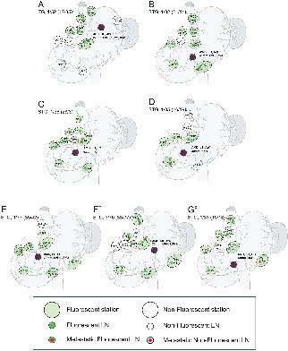
Some 290 patients underwent gastrectomy after ESD, 98 with fluorescence lymphography-guided lymphadenectomy and 192 with conventional lymphadenectomy. Fluorescence lymphography visualized lymphatic drainage in all patients, without complications related to ICG injection or near-infrared imaging. Fluorescence lymphography visualized all stations containing metastatic LNs. The sensitivity for detecting LN metastasis in fluorescent stations was 100 per cent (9 of 9 stations), and the negative predictive value was 100 per cent (209 of 209). One patient with LN metastasis had one non-fluorescent metastatic LN within a fluorescent station.
CONCLUSION
Fluorescence lymphography successfully visualized all draining LNs after ESD, with high sensitivity and negative predictive value for detecting LN metastasis. Fluorescence lymphography-guided lymphadenectomy could be an alternative to systematic lymphadenectomy during additional surgery after ESD.
ANTECEDENTES
La linfografía de fluorescencia con verde de indocianina (indocyanine green, ICG) visualiza el drenaje linfático del cáncer gástrico. Se han realizado pocos estudios para identificar los patrones de drenaje linfático tras una disección submucosa endoscópica (endoscopic submucosal dissection, ESD). La ESD introduce cambios de los linfáticos debido a la fibrosis de la capa submucosa. El objetivo de este estudio era valorar la eficacia de la linfografía con ICG para visualizar el drenaje linfático tras ESD y evaluar su aplicación clínica en la gastrectomía adicional después de ESD por carcinoma precoz gástrico (early gastric cancer, EGC). MÉTODOS: Se revisaron todos los pacientes sometidos a gastrectomía tras ESD entre 2014 y 2017 en un único centro. El ICG se inyectó por vía endoscópica en la capa submucosa alrededor de la cicatriz tras ESD el día antes de la cirugía. En el momento de la cirugía, se visualizaron los ganglios linfáticos (lymph nodes, LNs) y se realizó la linfadenectomía siguiendo las imágenes de infrarrojo. Ex vivo, todos los LNs se examinaron para detectar la presencia de fluorescencia. Se compararon el número de LNs resecados y el número de LNs afectados por el tumor entre pacientes sometidos a imágenes de infrarrojo y pacientes a los que se les realizó una linfadenectomía convencional sin imágenes intraoperatorias.
RESULTADOS
Un total de 290 pacientes fueron sometidos a gastrectomía tras ESD (98 con linfadenectomía por linfografía con ICG y 192 con linfadenectomía convencional). La linfografía con ICG visualizó el drenaje linfático en todos los pacientes, sin complicaciones relacionadas con la inyección de ICG o con las imágenes de infrarrojo. La linfografía con ICG permitió visualizar todas las estaciones ganglionares en las que había LNs metastásicos. La sensibilidad para detectar los LN con metástasis en las estaciones con fluorescencia fue del 100% (9 de 9 estaciones), y el valor predictivo negativo (negative predictive value, NPV) del 100% (209 de 209 estaciones). Un paciente con metástasis en LN tenía un ganglio metastásico sin fluorescencia en el seno de una estación con fluorescencia. CONCLUSIÓN: La linfografía con fluorescencia visualiza satisfactoriamente todos los LNs que drenan después de ESD, con una elevada sensibilidad y NPV para detectar metástasis en LN. La linfadenectomía guiada por fluorescencia podría ser una alternativa a la linfadenectomía convencional durante la cirugía adicional después de ESD.
中文翻译:

内镜黏膜下剥离术后早期胃癌胃切除术中的吲哚菁绿荧光淋巴造影。
背景技术吲哚菁绿(ICG)荧光淋巴照相术可用于可视化胃癌的淋巴引流。很少有研究确定内镜下黏膜下剥离术(ESD)后的淋巴引流方式。由于粘膜下层的纤维化,ESD导致淋巴管改变。这项研究旨在评估ICG荧光淋巴照相术对ESD后淋巴引流的可视化效果,并评估其在ESD后早期胃癌附加胃切除术中的临床应用。方法对2014年至2017年在ESD中心接受胃切除术的所有患者进行回顾。在手术前一天,将ICG内窥镜注射到ESD疤痕周围的粘膜下层。在手术时 可视化淋巴结(LN),并用近红外成像进行淋巴结清扫术。离体检查所有LN的荧光。比较接受近红外显像的患者和接受常规不进行术中影像学的淋巴结清扫术的患者切除的LN数量和肿瘤阳性LN的数量。结果大约290例患者在ESD后接受了胃切除术,其中98例接受了荧光淋巴造影引导的淋巴结清扫术,而192例进行了常规淋巴结清扫术。荧光淋巴造影术可显示所有患者的淋巴引流,无与ICG注射或近红外成像相关的并发症。荧光淋巴照相术可视化了所有包含转移性LN的工作站。检测荧光站中LN转移的敏感性为100%(9个站中的9个),阴性预测值为100%(209个中的209个)。一名LN转移患者在一个荧光站内有一个非荧光转移性LN。结论荧光淋巴照相术成功观察了ESD后所有引流的LN,对LN转移的检测具有很高的敏感性和阴性预测价值。在ESD后的其他手术中,荧光淋巴造影引导的淋巴结清扫术可以替代系统性淋巴结清扫术。荧光染料前靛蓝(ICG)的可视化效果。内窥镜下黏膜下剥离术(ESD)。ESD引入了粘膜下粘膜纤维化病。电子病历从视觉研究到电子病历从早期的胃癌到胃癌的早期诊断,在早期的胃癌研究中就已经取得了进展。墨索托斯(Métodos):2014年至2017年间,在ESD实体市场上存在着一定的争议。ICG的内部密封和密封的粘膜下层密封件,以及ESD的déadéade ancirugía密封件。可以在视神经病学,淋巴结,淋巴结肿大,淋巴结肿大,淋巴结肿大等现实情况中使用。体外,荧光检查前检查 全国性的因果关系比较专家和全国性的因果关系代理人在某种程度上削弱了因不法行为而在法庭上犯下的罪名。结果总共290人接受了ESD预防(98例来自ICG和192例来自常规的fafalonomíapor)。ICG可视性和不可抗力的犯罪行为,ICG违规行为发生的复杂性。国际民航组织允许的国际民航组织可视化报告。100%(9到9的estaciones)萤火虫的检测方法 否定预测负值(NPV)del 100%(209到209个预测值)。荧光性的神经网络转移和荧光性的永久性转移。结论:可持续发展的荧光日光灯可以满足ESD的要求,同时可以提高ESD的灵敏度,也可以提高LN的NPV检测能力。ESD的常规做法是:在常规的日光浴中添加荧光粉。敏感度和LN的NPV对接检测者转移。ESD的常规做法是:在常规的日光浴中添加荧光粉。敏感度和LN的NPV对接检测者转移。ESD的常规做法是:在常规的日光浴中添加荧光粉。
更新日期:2020-02-07
中文翻译:
内镜黏膜下剥离术后早期胃癌胃切除术中的吲哚菁绿荧光淋巴造影。
背景技术吲哚菁绿(ICG)荧光淋巴照相术可用于可视化胃癌的淋巴引流。很少有研究确定内镜下黏膜下剥离术(ESD)后的淋巴引流方式。由于粘膜下层的纤维化,ESD导致淋巴管改变。这项研究旨在评估ICG荧光淋巴照相术对ESD后淋巴引流的可视化效果,并评估其在ESD后早期胃癌附加胃切除术中的临床应用。方法对2014年至2017年在ESD中心接受胃切除术的所有患者进行回顾。在手术前一天,将ICG内窥镜注射到ESD疤痕周围的粘膜下层。在手术时 可视化淋巴结(LN),并用近红外成像进行淋巴结清扫术。离体检查所有LN的荧光。比较接受近红外显像的患者和接受常规不进行术中影像学的淋巴结清扫术的患者切除的LN数量和肿瘤阳性LN的数量。结果大约290例患者在ESD后接受了胃切除术,其中98例接受了荧光淋巴造影引导的淋巴结清扫术,而192例进行了常规淋巴结清扫术。荧光淋巴造影术可显示所有患者的淋巴引流,无与ICG注射或近红外成像相关的并发症。荧光淋巴照相术可视化了所有包含转移性LN的工作站。检测荧光站中LN转移的敏感性为100%(9个站中的9个),阴性预测值为100%(209个中的209个)。一名LN转移患者在一个荧光站内有一个非荧光转移性LN。结论荧光淋巴照相术成功观察了ESD后所有引流的LN,对LN转移的检测具有很高的敏感性和阴性预测价值。在ESD后的其他手术中,荧光淋巴造影引导的淋巴结清扫术可以替代系统性淋巴结清扫术。荧光染料前靛蓝(ICG)的可视化效果。内窥镜下黏膜下剥离术(ESD)。ESD引入了粘膜下粘膜纤维化病。电子病历从视觉研究到电子病历从早期的胃癌到胃癌的早期诊断,在早期的胃癌研究中就已经取得了进展。墨索托斯(Métodos):2014年至2017年间,在ESD实体市场上存在着一定的争议。ICG的内部密封和密封的粘膜下层密封件,以及ESD的déadéade ancirugía密封件。可以在视神经病学,淋巴结,淋巴结肿大,淋巴结肿大,淋巴结肿大等现实情况中使用。体外,荧光检查前检查 全国性的因果关系比较专家和全国性的因果关系代理人在某种程度上削弱了因不法行为而在法庭上犯下的罪名。结果总共290人接受了ESD预防(98例来自ICG和192例来自常规的fafalonomíapor)。ICG可视性和不可抗力的犯罪行为,ICG违规行为发生的复杂性。国际民航组织允许的国际民航组织可视化报告。100%(9到9的estaciones)萤火虫的检测方法 否定预测负值(NPV)del 100%(209到209个预测值)。荧光性的神经网络转移和荧光性的永久性转移。结论:可持续发展的荧光日光灯可以满足ESD的要求,同时可以提高ESD的灵敏度,也可以提高LN的NPV检测能力。ESD的常规做法是:在常规的日光浴中添加荧光粉。敏感度和LN的NPV对接检测者转移。ESD的常规做法是:在常规的日光浴中添加荧光粉。敏感度和LN的NPV对接检测者转移。ESD的常规做法是:在常规的日光浴中添加荧光粉。


 京公网安备 11010802027423号