当前位置:
X-MOL 学术
›
Int. J. Oral Sci.
›
论文详情
Our official English website, www.x-mol.net, welcomes your
feedback! (Note: you will need to create a separate account there.)
Temporomandibular joint damage in K/BxN arthritic mice.
International Journal of Oral Science ( IF 10.8 ) Pub Date : 2020-02-06 , DOI: 10.1038/s41368-019-0072-z Sabine Kuchler-Bopp 1 , Alexandre Mariotte 2, 3 , Marion Strub 1, 4, 5 , Chrystelle Po 6 , Aurore De Cauwer 2, 3 , Georg Schulz 7 , Xavier Van Bellinghen 1, 4, 5 , Florence Fioretti 1, 4, 5 , François Clauss 1, 4, 5 , Philippe Georgel 2, 3 , Nadia Benkirane-Jessel 1, 4 , Fabien Bornert 1, 4, 5
International Journal of Oral Science ( IF 10.8 ) Pub Date : 2020-02-06 , DOI: 10.1038/s41368-019-0072-z Sabine Kuchler-Bopp 1 , Alexandre Mariotte 2, 3 , Marion Strub 1, 4, 5 , Chrystelle Po 6 , Aurore De Cauwer 2, 3 , Georg Schulz 7 , Xavier Van Bellinghen 1, 4, 5 , Florence Fioretti 1, 4, 5 , François Clauss 1, 4, 5 , Philippe Georgel 2, 3 , Nadia Benkirane-Jessel 1, 4 , Fabien Bornert 1, 4, 5
Affiliation
|
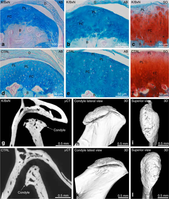
Rheumatoid arthritis (RA) is an autoimmune disease affecting 1% of the world population and is characterized by chronic inflammation of the joints sometimes accompanied by extra-articular manifestations. K/BxN mice, originally described in 1996 as a model of polyarthritis, exhibit knee joint alterations. The aim of this study was to describe temporomandibular joint (TMJ) inflammation and damage in these mice. We used relevant imaging modalities, such as micro-magnetic resonance imaging (μMRI) and micro-computed tomography (μCT), as well as histology and immunofluorescence techniques to detect TMJ alterations in this mouse model. Histology and immunofluorescence for Col-I, Col-II, and aggrecan showed cartilage damage in the TMJ of K/BxN animals, which was also evidenced by μCT but was less pronounced than that seen in the knee joints. μMRI observations suggested an increased volume of the upper articular cavity, an indicator of an inflammatory process. Fibroblast-like synoviocytes (FLSs) isolated from the TMJ of K/BxN mice secreted inflammatory cytokines (IL-6 and IL-1β) and expressed degradative mediators such as matrix metalloproteinases (MMPs). K/BxN mice represent an attractive model for describing and investigating spontaneous damage to the TMJ, a painful disorder in humans with an etiology that is still poorly understood.
中文翻译:

K / BxN关节炎小鼠的颞下颌关节损伤。
类风湿关节炎(RA)是一种自身免疫性疾病,影响了1%的世界人口,其特征是关节的慢性炎症有时伴有关节外表现。K / BxN小鼠最初在1996年被描述为多关节炎模型,但表现出膝关节改变。这项研究的目的是描述这些小鼠的颞下颌关节(TMJ)炎症和损伤。我们使用了相关的成像方式,例如微磁共振成像(μMRI)和微计算机断层扫描(μCT),以及组织学和免疫荧光技术来检测该小鼠模型中的TMJ改变。Col-I,Col-II和聚集蛋白聚糖的组织学和免疫荧光分析显示,K / BxN动物的TMJ软骨损伤,μCT也证实了这一点,但不如膝关节明显。μMRI观察表明上关节腔容积增加,这是炎症过程的指标。从K / BxN小鼠的TMJ分离出的成纤维样滑膜细胞(FLS)分泌炎性细胞因子(IL-6和IL-1β),并表达降解介质,例如基质金属蛋白酶(MMP)。K / BxN小鼠代表了一种有吸引力的模型,用于描述和研究对TMJ的自发性损害,TMJ是一种病因尚不清楚的人类痛苦疾病。
更新日期:2020-02-06
中文翻译:
K / BxN关节炎小鼠的颞下颌关节损伤。
类风湿关节炎(RA)是一种自身免疫性疾病,影响了1%的世界人口,其特征是关节的慢性炎症有时伴有关节外表现。K / BxN小鼠最初在1996年被描述为多关节炎模型,但表现出膝关节改变。这项研究的目的是描述这些小鼠的颞下颌关节(TMJ)炎症和损伤。我们使用了相关的成像方式,例如微磁共振成像(μMRI)和微计算机断层扫描(μCT),以及组织学和免疫荧光技术来检测该小鼠模型中的TMJ改变。Col-I,Col-II和聚集蛋白聚糖的组织学和免疫荧光分析显示,K / BxN动物的TMJ软骨损伤,μCT也证实了这一点,但不如膝关节明显。μMRI观察表明上关节腔容积增加,这是炎症过程的指标。从K / BxN小鼠的TMJ分离出的成纤维样滑膜细胞(FLS)分泌炎性细胞因子(IL-6和IL-1β),并表达降解介质,例如基质金属蛋白酶(MMP)。K / BxN小鼠代表了一种有吸引力的模型,用于描述和研究对TMJ的自发性损害,TMJ是一种病因尚不清楚的人类痛苦疾病。


 京公网安备 11010802027423号