当前位置:
X-MOL 学术
›
Biotechnol. Bioeng.
›
论文详情
Our official English website, www.x-mol.net, welcomes your
feedback! (Note: you will need to create a separate account there.)
ECM concentration and cell-mediated traction forces play a role in vascular network assembly in 3D bioprinted tissue.
Biotechnology and Bioengineering ( IF 3.5 ) Pub Date : 2020-01-11 , DOI: 10.1002/bit.27250 Guangliang Zhang 1, 2 , Mathew Varkey 1 , Zhan Wang 1 , Beibei Xie 1, 2 , Ruixing Hou 2 , Anthony Atala 1
Biotechnology and Bioengineering ( IF 3.5 ) Pub Date : 2020-01-11 , DOI: 10.1002/bit.27250 Guangliang Zhang 1, 2 , Mathew Varkey 1 , Zhan Wang 1 , Beibei Xie 1, 2 , Ruixing Hou 2 , Anthony Atala 1
Affiliation
|
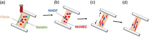
Tissue vascularization is critical to enable oxygen and nutrient supply. Therefore, establishing expedient vasculature is necessary for the survival of tissue after transplantation. The use of biomechanical forces, such as cell‐induced traction forces, may be a promising method to encourage growth of the vascular network. Three‐dimensional (3D) bioprinting, which offers unprecedented versatility through precise control over spatial distribution and structure of tissue constructs, can be used to generate capillary‐like structures in vitro that would mimic microvessels. This study aimed to develop an in vitro, 3D bioprinted tissue model to study the effect of cellular forces on the spatial organization of vascular structures and tissue maturation. The developed in vitro model consists of a 3D bioprinted polycaprolactone (PCL) frame with a gelatin spacer hydrogel layer and a gelatin–fibrin–hyaluronic acid hydrogel layer containing normal human dermal fibroblasts and human umbilical vein endothelial cells printed as vessel lines on top. The formation of vessel‐like networks and vessel lumens in the 3D bioprinted in vitro model was assessed at different fibrinogen concentrations with and without inhibitors of cell‐mediated traction forces. Constructs containing 5 mg/ml fibrinogen had longer vessels compared to the other concentrations of fibrinogen used. Also, for all concentrations of fibrinogen used, most of the vessel‐like structures grew parallel to the direction the PCL frame‐mediated tensile forces, with very few branching structures observed. Treatment of the 3D bioprinted constructs with traction inhibitors resulted in a significant reduction in length of vessel‐like networks. The 3D bioprinted constructs also had better lumen formation, increased collagen deposition, more elaborate actin networks, and well‐aligned matrix fibers due to the increased cell‐mediated traction forces present compared to the non‐anchored, floating control constructs. This study showed that cell traction forces from the actomyosin complex are critical for vascular network assembly in 3D bioprinted tissue. Strategies involving the use of cell‐mediated traction forces may be promising for the development of bioprinting approaches for fabrication of vascularized tissue constructs.
中文翻译:

ECM浓度和细胞介导的牵引力在3D生物打印组织中的血管网络组装中起作用。
组织血管形成对于确保氧气和营养供应至关重要。因此,建立合适的脉管系统对于移植后组织的存活是必要的。使用生物力学力,例如细胞诱导的牵引力,可能是鼓励血管网络生长的有前途的方法。三维(3D)生物打印通过精确控制组织结构的空间分布和结构而提供了前所未有的多功能性,可用于在体外生成类似于微血管的毛细管状结构。这项研究旨在开发体外3D生物打印的组织模型,以研究细胞力对血管结构的空间组织和组织成熟的影响。开发的体外模型由3D生物打印的聚己内酯(PCL)框架组成,明胶间隔物水凝胶层和明胶-纤维蛋白-透明质酸水凝胶层包含正常人皮肤成纤维细胞和人脐静脉内皮细胞,上面印有血管线。在有或没有细胞介导的牵引力抑制剂的情况下,在不同的纤维蛋白原浓度下,评估了3D生物打印体外模型中血管样网络和血管腔的形成。与使用的其他浓度的纤维蛋白原相比,含有5 mg / ml纤维蛋白原的构建体具有更长的血管。同样,对于所有浓度的纤维蛋白原,大多数血管样结构都平行于PCL框架介导的拉力方向生长,观察到的分支结构很少。用牵引抑制剂处理3D生物打印的结构可显着减少血管样网络的长度。与非锚定的浮动对照构建体相比,由于存在增加的细胞介导的牵引力,因此3D生物打印的构建体还具有更好的内腔形成,增加的胶原蛋白沉积,更精细的肌动蛋白网络以及排列良好的基质纤维。这项研究表明,来自肌动球蛋白复合物的细胞牵引力对于3D生物打印组织中的血管网络组装至关重要。涉及使用细胞介导的牵引力的策略可能对开发用于制造血管化组织构造的生物打印方法很有前途。与非锚定的浮动对照构建体相比,由于存在增加的细胞介导的牵引力,因此3D生物打印的构建体还具有更好的内腔形成,增加的胶原蛋白沉积,更精细的肌动蛋白网络以及排列良好的基质纤维。这项研究表明,来自肌动球蛋白复合物的细胞牵引力对于3D生物打印组织中的血管网络组装至关重要。涉及使用细胞介导的牵引力的策略可能对开发用于制造血管化组织构造的生物打印方法很有前途。与非锚定的浮动对照构建体相比,由于存在增加的细胞介导的牵引力,因此3D生物打印的构建体还具有更好的内腔形成,增加的胶原蛋白沉积,更精细的肌动蛋白网络以及排列良好的基质纤维。这项研究表明,来自肌动球蛋白复合物的细胞牵引力对于3D生物打印组织中的血管网络组装至关重要。涉及使用细胞介导的牵引力的策略可能对开发用于制造血管化组织构造的生物打印方法很有前途。这项研究表明,来自肌动球蛋白复合物的细胞牵引力对于3D生物打印组织中的血管网络组装至关重要。涉及使用细胞介导的牵引力的策略可能对开发用于制造血管化组织构造的生物打印方法很有前途。这项研究表明,来自肌动球蛋白复合物的细胞牵引力对于3D生物打印组织中的血管网络组装至关重要。涉及使用细胞介导的牵引力的策略可能对开发用于制造血管化组织构造的生物打印方法很有前途。
更新日期:2020-03-09
中文翻译:
ECM浓度和细胞介导的牵引力在3D生物打印组织中的血管网络组装中起作用。
组织血管形成对于确保氧气和营养供应至关重要。因此,建立合适的脉管系统对于移植后组织的存活是必要的。使用生物力学力,例如细胞诱导的牵引力,可能是鼓励血管网络生长的有前途的方法。三维(3D)生物打印通过精确控制组织结构的空间分布和结构而提供了前所未有的多功能性,可用于在体外生成类似于微血管的毛细管状结构。这项研究旨在开发体外3D生物打印的组织模型,以研究细胞力对血管结构的空间组织和组织成熟的影响。开发的体外模型由3D生物打印的聚己内酯(PCL)框架组成,明胶间隔物水凝胶层和明胶-纤维蛋白-透明质酸水凝胶层包含正常人皮肤成纤维细胞和人脐静脉内皮细胞,上面印有血管线。在有或没有细胞介导的牵引力抑制剂的情况下,在不同的纤维蛋白原浓度下,评估了3D生物打印体外模型中血管样网络和血管腔的形成。与使用的其他浓度的纤维蛋白原相比,含有5 mg / ml纤维蛋白原的构建体具有更长的血管。同样,对于所有浓度的纤维蛋白原,大多数血管样结构都平行于PCL框架介导的拉力方向生长,观察到的分支结构很少。用牵引抑制剂处理3D生物打印的结构可显着减少血管样网络的长度。与非锚定的浮动对照构建体相比,由于存在增加的细胞介导的牵引力,因此3D生物打印的构建体还具有更好的内腔形成,增加的胶原蛋白沉积,更精细的肌动蛋白网络以及排列良好的基质纤维。这项研究表明,来自肌动球蛋白复合物的细胞牵引力对于3D生物打印组织中的血管网络组装至关重要。涉及使用细胞介导的牵引力的策略可能对开发用于制造血管化组织构造的生物打印方法很有前途。与非锚定的浮动对照构建体相比,由于存在增加的细胞介导的牵引力,因此3D生物打印的构建体还具有更好的内腔形成,增加的胶原蛋白沉积,更精细的肌动蛋白网络以及排列良好的基质纤维。这项研究表明,来自肌动球蛋白复合物的细胞牵引力对于3D生物打印组织中的血管网络组装至关重要。涉及使用细胞介导的牵引力的策略可能对开发用于制造血管化组织构造的生物打印方法很有前途。与非锚定的浮动对照构建体相比,由于存在增加的细胞介导的牵引力,因此3D生物打印的构建体还具有更好的内腔形成,增加的胶原蛋白沉积,更精细的肌动蛋白网络以及排列良好的基质纤维。这项研究表明,来自肌动球蛋白复合物的细胞牵引力对于3D生物打印组织中的血管网络组装至关重要。涉及使用细胞介导的牵引力的策略可能对开发用于制造血管化组织构造的生物打印方法很有前途。这项研究表明,来自肌动球蛋白复合物的细胞牵引力对于3D生物打印组织中的血管网络组装至关重要。涉及使用细胞介导的牵引力的策略可能对开发用于制造血管化组织构造的生物打印方法很有前途。这项研究表明,来自肌动球蛋白复合物的细胞牵引力对于3D生物打印组织中的血管网络组装至关重要。涉及使用细胞介导的牵引力的策略可能对开发用于制造血管化组织构造的生物打印方法很有前途。


 京公网安备 11010802027423号