Journal of Neuroradiology ( IF 3.0 ) Pub Date : 2019-12-13 , DOI: 10.1016/j.neurad.2019.11.006 Fumio Suzuki 1 , Noriko Sato 2 , Atsuhiko Sugiyama 3 , Keiya Iijima 4 , Yoko Shigemoto 2 , Emiko Morimoto 2 , Yukio Kimura 2 , Hiroyuki Fujii 2 , Yuji Takahashi 5 , Yasuhiro Nakata 6 , Hiroshi Matsuda 7 , Osamu Abe 8
|
Background and purpose
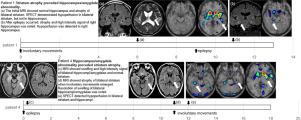
Chorea-acanthocytosis, a rare neurodegenerative disease, affects both the striatum and the medial temporal lobe which may cause involuntary movements and epilepsy, respectively. We examined the imaging changes of the hippocampus/amygdala and the striatum as well as clinical symptoms.
Materials and methods
We retrospectively reviewed 29 MRI and 13 SPECT studies and the clinical findings of seven genetically confirmed chorea-acanthocytosis patients. We evaluated the time-dependent imaging changes of the hippocampus/amygdala and striatum and examined the relationships among these images and symptoms.
Results
The initial symptom was epilepsy in four patients and involuntary movements in three patients. These symptoms were eventually noted in five and all seven patients, respectively. On MRI, most patients showed striatum atrophy before a hippocampus/amygdala abnormality emerged, but one patient showed a hippocampus/amygdala abnormality before striatum atrophy. Abnormal MRI findings of hippocampus/amygdala were noted in five patients and atrophy of striatum in all seven patients. SPECT demonstrated hypoperfusion of hippocampus/amygdala in three patients and that of striatum in all five available patients. Four patients demonstrated hypoperfusion of striatum earlier than that of hippocampus/amygdala and one patient showed hypoperfusion of both simultaneously. Many imaging abnormal lesions were accompanied by their corresponding symptoms, but not always so.
Conclusion
Striatum abnormalities were the initial imaging findings in many chorea-acanthocytosis patients, but epilepsy or hippocampus/amygdala imaging abnormalities may be the only findings at the early stage. It is important to understand the detailed clinical and imaging time courses for the diagnosis of chorea-acanthocytosis.
中文翻译:
舞蹈病-棘红细胞增多症:症状和影像学表现的时间依赖性变化
背景和目的
舞蹈病-棘红细胞增多症是一种罕见的神经退行性疾病,影响纹状体和内侧颞叶,可能分别导致不自主运动和癫痫。我们检查了海马/杏仁核和纹状体的影像学变化以及临床症状。
材料和方法
我们回顾性审查了 29 项 MRI 和 13 项 SPECT 研究以及 7 名经基因证实的舞蹈病-棘红细胞增多症患者的临床发现。我们评估了海马/杏仁核和纹状体的时间依赖性成像变化,并检查了这些图像和症状之间的关系。
结果
最初的症状是四名患者的癫痫和三名患者的不自主运动。这些症状最终分别在 5 名和所有 7 名患者中出现。在MRI上,大多数患者在出现海马/杏仁核异常之前表现出纹状体萎缩,但一名患者在出现纹状体萎缩之前表现出海马/杏仁核异常。5 名患者的海马/杏仁核的 MRI 结果异常,所有 7 名患者均出现纹状体萎缩。SPECT 显示三名患者的海马/杏仁核灌注不足,所有五名可用患者的纹状体灌注不足。四名患者比海马/杏仁核更早地表现出纹状体的低灌注,一名患者同时表现出两者的低灌注。许多影像学异常病变伴随着相应的症状,
结论
纹状体异常是许多舞蹈病-棘红细胞增多症患者的最初影像学表现,但癫痫或海马/杏仁核影像学异常可能是早期唯一的发现。了解详细的临床和影像学时程对于舞蹈病-棘红细胞增多症的诊断很重要。


 京公网安备 11010802027423号