Current Pharmaceutical Biotechnology ( IF 2.2 ) Pub Date : 2020-05-31 , DOI: 10.2174/1389201021666191224121206 Magdalena Skonieczna 1, 2 , Malgorzata Adamiec 1, 2 , Dorota Hudy 1, 2 , Patrycja Nieslon 3 , Daniel Fochtman 3 , Patryk Bil 1, 2
|
Background: Changes in the cellular behavior depend on environmental and intracellular interactions. Cancer treatments force the changes, first on the molecular level, but the main visible changes are macroscopic. During radiotherapy, cancer cell’s adhesion, proliferation and migration should be well monitored. In over 60% of diagnosed cancers cases, patients are given treatments with different protocols of radiotherapy, which result in possible metastasis and acute whole body response to toxic radiation.
Objective: Effectiveness of the therapy used depends on the sensitivity/resistance of irradiated cancer cells. Cellular mechanisms of cancer protection, such as the activation of DNA damage and repair pathways, antioxidants production and oxidative stress suppression during treatments are not desirable. Cancer cells monitoring require the development of novel techniques, and the best techniques are non-invasive and long-term live observation methods, which are shown in this study.
Methods: In cancers, invasive and metastatic phenotypes could be enhanced by stimulation of proliferation rate, decreased adhesion with simultaneous increase of motility and migration potential. For such reasons, the Ionizing Radiation (IR) stimulated proliferation; migration with lowered adhesiveness of cancer Me45 and normal fibroblasts NHDF were studied. Using impedance measurements technique for live cells, the adhesion of cells after IR exposition was assessed. Additionally proliferation and migration potential, based on standard Wound Healing assay were evaluated by timelapse microscopic observations.
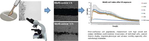
Results: We found simulative IR dose-ranges (0.2-2 Gy) for Me45 and NHDF cells, with higher proliferation and adhesion rates. On the other hand, lethal impact of IR (10-12 Gy) on both the cell lines was indicated.
Conclusion: Over-confluence cell populations, characterized with high crowd and contact inhibition could modulate invasiveness of individual cells, convert them to display migration phenotype and advance motility, especially after radiotherapy treatments.
中文翻译:
电离辐射后细胞粘附,增殖和迁移的活阻抗测量和延时显微镜观察。
背景:细胞行为的变化取决于环境和细胞内的相互作用。癌症治疗首先在分子水平上迫使这种改变,但是主要的可见改变是宏观的。在放疗期间,应密切监测癌细胞的粘附,增殖和迁移。在超过60%的确诊癌症病例中,为患者提供了不同放疗方案的治疗,这可能导致转移和对毒性辐射的急性全身反应。
目的:所用疗法的有效性取决于放射癌细胞的敏感性/耐药性。癌症保护的细胞机制,例如DNA损伤的激活和修复途径,抗氧化剂的产生以及治疗期间的氧化应激抑制,是不理想的。癌细胞监测需要开发新技术,而最佳技术是非侵入性和长期的实时观察方法,本研究显示。
方法:在癌症中,可以通过刺激增殖率,减少粘附并同时增加运动性和迁移潜能来增强侵袭性和转移性表型。由于这些原因,电离辐射(IR)刺激了增殖;研究了在癌症Me45和正常成纤维细胞NHDF黏附力降低的情况下的迁移。使用针对活细胞的阻抗测量技术,评估了IR暴露后细胞的粘附力。另外,通过延时显微镜观察评估了基于标准伤口愈合测定的增殖和迁移潜力。
结果:我们发现Me45和NHDF细胞的模拟IR剂量范围(0.2-2 Gy),具有更高的增殖和粘附率。另一方面,表明了IR(10-12 Gy)对两种细胞系的致命影响。
结论:以高人群和接触抑制为特征的过度汇合的细胞群可以调节单个细胞的侵袭性,使其转化为迁移表型并促进运动,特别是在放疗后。


 京公网安备 11010802027423号