Our official English website, www.x-mol.net, welcomes your
feedback! (Note: you will need to create a separate account there.)
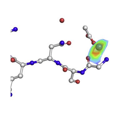
Experimental Phasing of MicroED Data Using Radiation Damage.
Structure ( IF 4.4 ) Pub Date : 2020-02-04 , DOI: 10.1016/j.str.2020.01.008 Michael W Martynowycz 1 , Johan Hattne 1 , Tamir Gonen 1
Structure ( IF 4.4 ) Pub Date : 2020-02-04 , DOI: 10.1016/j.str.2020.01.008 Michael W Martynowycz 1 , Johan Hattne 1 , Tamir Gonen 1
Affiliation
|
We previously demonstrated that microcrystal electron diffraction (MicroED) can be used to determine atomic-resolution structures from vanishingly small three-dimensional crystals. Here, we present an example of an experimentally phased structure using only MicroED data. The structure of a seven-residue peptide is solved starting from differences to the diffraction intensities induced by structural changes due to radiation damage. The same wedge of reciprocal space was recorded twice by continuous-rotation MicroED from a set of 11 individual crystals. The data from the first pass were merged to make a "low-dose dataset." The data from the second pass were similarly merged to form a "damaged dataset." Differences between these two datasets were used to identify a single heavy-atom site from a Patterson difference map, and initial phases were generated. Finally, the structure was completed by iterative cycles of modeling and refinement.
中文翻译:

使用辐射损伤对 MicroED 数据进行实验定相。
我们之前证明,微晶电子衍射 (MicroED) 可用于从极小的三维晶体中确定原子分辨率结构。在这里,我们展示了一个仅使用 MicroED 数据的实验阶段结构示例。解决了七残基肽的结构,从辐射损伤引起的结构变化引起的衍射强度差异开始。连续旋转的 MicroED 从一组 11 个单独的晶体中记录了两次倒易空间的相同楔形。来自第一遍的数据被合并成一个“低剂量数据集”。来自第二遍的数据被类似地合并形成一个“损坏的数据集”。这两个数据集之间的差异用于从帕特森差异图中识别单个重原子位点,并生成了初始阶段。最后,通过建模和细化的迭代循环完成结构。
更新日期:2020-02-04
中文翻译:
使用辐射损伤对 MicroED 数据进行实验定相。
我们之前证明,微晶电子衍射 (MicroED) 可用于从极小的三维晶体中确定原子分辨率结构。在这里,我们展示了一个仅使用 MicroED 数据的实验阶段结构示例。解决了七残基肽的结构,从辐射损伤引起的结构变化引起的衍射强度差异开始。连续旋转的 MicroED 从一组 11 个单独的晶体中记录了两次倒易空间的相同楔形。来自第一遍的数据被合并成一个“低剂量数据集”。来自第二遍的数据被类似地合并形成一个“损坏的数据集”。这两个数据集之间的差异用于从帕特森差异图中识别单个重原子位点,并生成了初始阶段。最后,通过建模和细化的迭代循环完成结构。


 京公网安备 11010802027423号