当前位置:
X-MOL 学术
›
Nat. Protoc.
›
论文详情
Our official English website, www.x-mol.net, welcomes your
feedback! (Note: you will need to create a separate account there.)
Multicolor two-photon imaging of in vivo cellular pathophysiology upon influenza virus infection using the two-photon IMPRESS.
Nature Protocols ( IF 13.1 ) Pub Date : 2020-01-29 , DOI: 10.1038/s41596-019-0275-y Hiroshi Ueki 1 , I-Hsuan Wang 1 , Dongming Zhao 1, 2 , Matthias Gunzer 3 , Yoshihiro Kawaoka 1, 4, 5
Nature Protocols ( IF 13.1 ) Pub Date : 2020-01-29 , DOI: 10.1038/s41596-019-0275-y Hiroshi Ueki 1 , I-Hsuan Wang 1 , Dongming Zhao 1, 2 , Matthias Gunzer 3 , Yoshihiro Kawaoka 1, 4, 5
Affiliation
|
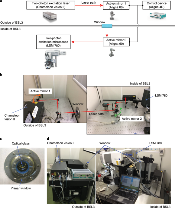
In vivo two-photon imaging is a valuable technique for studies of viral pathogenesis and host responses to infection in vivo. In this protocol, we describe a methodology for analyzing influenza virus-infected lung in vivo by two-photon imaging microscopy. We describe the surgical procedure, how to stabilize the lung, and an approach to analyzing the data. Further, we provide a database of fluorescent dyes, antibodies, and reporter mouse lines that can be used in combination with a reporter influenza virus (Color-flu) for multicolor analysis. Setup of this model typically takes ~30 min and enables the observation of influenza virus-infected lungs for >4 h during the acute phase of the inflammation and at least 1 h in the lethal phase. This imaging system, which we termed two-photon IMPRESS (imaging pathophysiology research system), is broadly applicable to analyses of other respiratory pathogens and reveals disease progression at the cellular level in vivo.
中文翻译:

使用双光子 IMPRESS 对流感病毒感染时的体内细胞病理生理学进行多色双光子成像。
体内双光子成像是研究病毒发病机制和宿主对体内感染反应的宝贵技术。在本协议中,我们描述了一种通过双光子成像显微镜分析体内流感病毒感染的肺部的方法。我们描述了手术过程、如何稳定肺部以及分析数据的方法。此外,我们还提供荧光染料、抗体和报告小鼠系的数据库,可与报告流感病毒(Color-flu)结合使用进行多色分析。该模型的建立通常需要约 30 分钟,并且能够在炎症急性期观察流感病毒感染的肺部 >4 小时,在致死期至少观察 1 小时。该成像系统被我们称为双光子 IMPRESS(成像病理生理学研究系统),广泛适用于其他呼吸道病原体的分析,并揭示体内细胞水平的疾病进展。
更新日期:2020-01-29
中文翻译:
使用双光子 IMPRESS 对流感病毒感染时的体内细胞病理生理学进行多色双光子成像。
体内双光子成像是研究病毒发病机制和宿主对体内感染反应的宝贵技术。在本协议中,我们描述了一种通过双光子成像显微镜分析体内流感病毒感染的肺部的方法。我们描述了手术过程、如何稳定肺部以及分析数据的方法。此外,我们还提供荧光染料、抗体和报告小鼠系的数据库,可与报告流感病毒(Color-flu)结合使用进行多色分析。该模型的建立通常需要约 30 分钟,并且能够在炎症急性期观察流感病毒感染的肺部 >4 小时,在致死期至少观察 1 小时。该成像系统被我们称为双光子 IMPRESS(成像病理生理学研究系统),广泛适用于其他呼吸道病原体的分析,并揭示体内细胞水平的疾病进展。


 京公网安备 11010802027423号