Our official English website, www.x-mol.net, welcomes your
feedback! (Note: you will need to create a separate account there.)
Controlled clustering enhances PDX1 and NKX6.1 expression in pancreatic endoderm cells derived from pluripotent stem cells.
Scientific Reports ( IF 3.8 ) Pub Date : 2020-01-27 , DOI: 10.1038/s41598-020-57787-0 Raymond Tran 1 , Christopher Moraes 1, 2, 3 , Corinne A Hoesli 1, 2
Scientific Reports ( IF 3.8 ) Pub Date : 2020-01-27 , DOI: 10.1038/s41598-020-57787-0 Raymond Tran 1 , Christopher Moraes 1, 2, 3 , Corinne A Hoesli 1, 2
Affiliation
|
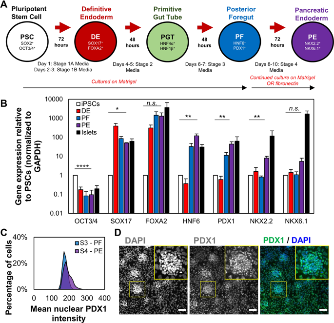
Pluripotent stem cell (PSC)-derived insulin-producing cells are a promising cell source for diabetes cellular therapy. However, the efficiency of the multi-step process required to differentiate PSCs towards pancreatic beta cells is variable between cell lines, batches and even within cultures. In adherent pancreatic differentiation protocols, we observed spontaneous local clustering of cells expressing elevated nuclear expression of pancreatic endocrine transcription factors, PDX1 and NKX6.1. Since aggregation has previously been shown to promote downstream differentiation, this local clustering may contribute to the variability in differentiation efficiencies observed within and between cultures. We therefore hypothesized that controlling and directing the spontaneous clustering process would lead to more efficient and consistent induction of pancreatic endocrine fate. Micropatterning cells in adherent microwells prompted clustering, local cell density increases, and increased nuclear accumulation of PDX1 and NKX6.1. Improved differentiation profiles were associated with distinct filamentous actin architectures, suggesting a previously overlooked role for cell-driven morphogenetic changes in supporting pancreatic differentiation. This work demonstrates that confined differentiation in cell-adhesive micropatterns may provide a facile, scalable, and more reproducible manufacturing route to drive morphogenesis and produce well-differentiated pancreatic cell clusters.
中文翻译:

受控聚类增强了多能干细胞来源的胰腺内胚层细胞中PDX1和NKX6.1的表达。
多能干细胞(PSC)衍生的胰岛素产生细胞是糖尿病细胞治疗的有希望的细胞来源。但是,区分PSC向胰腺β细胞所需的多步骤过程的效率在细胞系,批次甚至培养物中是可变的。在贴壁胰腺分化方案中,我们观察到了表达胰腺内分泌转录因子PDX1和NKX6.1核表达升高的细胞的自发局部聚集。由于之前已显示聚集会促进下游分化,所以这种局部聚类可能会导致在培养物中和培养物中观察到的分化效率发生变化。因此,我们假设控制和指导自发簇集过程将导致更有效,更一致地诱导胰腺内分泌命运。贴壁微孔中的微图案化细胞促使聚集,局部细胞密度增加,PDX1和NKX6.1的核积累增加。分化特征的改善与独特的丝状肌动蛋白结构有关,表明细胞驱动的形态发生变化在支持胰腺分化中被忽略的作用。这项工作表明,在细胞粘附性微模式中的有限分化可能提供一种简便,可扩展且可重现的制造途径,以驱动形态发生并产生分化良好的胰腺细胞簇。贴壁微孔中的微图案化细胞促使聚集,局部细胞密度增加,PDX1和NKX6.1的核积累增加。分化特征的改善与独特的丝状肌动蛋白结构有关,表明细胞驱动的形态发生变化在支持胰腺分化中被忽略的作用。这项工作表明,在细胞粘附微模式中的局限性分化可能提供一种简便,可扩展且可重现的制造途径,以驱动形态发生并产生分化良好的胰腺细胞簇。贴壁微孔中的微图案化细胞促使聚集,局部细胞密度增加,PDX1和NKX6.1的核积累增加。分化特征的改善与独特的丝状肌动蛋白结构有关,表明细胞驱动的形态发生变化在支持胰腺分化中被忽略的作用。这项工作表明,在细胞粘附微模式中的局限性分化可能提供一种简便,可扩展且可重现的制造途径,以驱动形态发生并产生分化良好的胰腺细胞簇。提示细胞驱动的形态发生变化在支持胰腺分化中的作用被人们忽略了。这项工作表明,在细胞粘附微模式中的局限性分化可能提供一种简便,可扩展且可重现的制造途径,以驱动形态发生并产生分化良好的胰腺细胞簇。提示细胞驱动的形态发生变化在支持胰腺分化中的作用先前被忽略。这项工作表明,在细胞粘附性微模式中的有限分化可能提供一种简便,可扩展且可重现的制造途径,以驱动形态发生并产生分化良好的胰腺细胞簇。
更新日期:2020-01-27
中文翻译:
受控聚类增强了多能干细胞来源的胰腺内胚层细胞中PDX1和NKX6.1的表达。
多能干细胞(PSC)衍生的胰岛素产生细胞是糖尿病细胞治疗的有希望的细胞来源。但是,区分PSC向胰腺β细胞所需的多步骤过程的效率在细胞系,批次甚至培养物中是可变的。在贴壁胰腺分化方案中,我们观察到了表达胰腺内分泌转录因子PDX1和NKX6.1核表达升高的细胞的自发局部聚集。由于之前已显示聚集会促进下游分化,所以这种局部聚类可能会导致在培养物中和培养物中观察到的分化效率发生变化。因此,我们假设控制和指导自发簇集过程将导致更有效,更一致地诱导胰腺内分泌命运。贴壁微孔中的微图案化细胞促使聚集,局部细胞密度增加,PDX1和NKX6.1的核积累增加。分化特征的改善与独特的丝状肌动蛋白结构有关,表明细胞驱动的形态发生变化在支持胰腺分化中被忽略的作用。这项工作表明,在细胞粘附性微模式中的有限分化可能提供一种简便,可扩展且可重现的制造途径,以驱动形态发生并产生分化良好的胰腺细胞簇。贴壁微孔中的微图案化细胞促使聚集,局部细胞密度增加,PDX1和NKX6.1的核积累增加。分化特征的改善与独特的丝状肌动蛋白结构有关,表明细胞驱动的形态发生变化在支持胰腺分化中被忽略的作用。这项工作表明,在细胞粘附微模式中的局限性分化可能提供一种简便,可扩展且可重现的制造途径,以驱动形态发生并产生分化良好的胰腺细胞簇。贴壁微孔中的微图案化细胞促使聚集,局部细胞密度增加,PDX1和NKX6.1的核积累增加。分化特征的改善与独特的丝状肌动蛋白结构有关,表明细胞驱动的形态发生变化在支持胰腺分化中被忽略的作用。这项工作表明,在细胞粘附微模式中的局限性分化可能提供一种简便,可扩展且可重现的制造途径,以驱动形态发生并产生分化良好的胰腺细胞簇。提示细胞驱动的形态发生变化在支持胰腺分化中的作用被人们忽略了。这项工作表明,在细胞粘附微模式中的局限性分化可能提供一种简便,可扩展且可重现的制造途径,以驱动形态发生并产生分化良好的胰腺细胞簇。提示细胞驱动的形态发生变化在支持胰腺分化中的作用先前被忽略。这项工作表明,在细胞粘附性微模式中的有限分化可能提供一种简便,可扩展且可重现的制造途径,以驱动形态发生并产生分化良好的胰腺细胞簇。


 京公网安备 11010802027423号