Our official English website, www.x-mol.net, welcomes your
feedback! (Note: you will need to create a separate account there.)
Podocyte autophagy is associated with foot process effacement and proteinuria in patients with minimal change nephrotic syndrome.
PLOS ONE ( IF 2.9 ) Pub Date : 2020-01-24 , DOI: 10.1371/journal.pone.0228337 Ayu Ogawa-Akiyama 1 , Hitoshi Sugiyama 2 , Masashi Kitagawa 1 , Keiko Tanaka 1, 3 , Yuzuki Kano 1 , Koki Mise 1 , Nozomu Otaka 2 , Katsuyuki Tanabe 1 , Hiroshi Morinaga 4 , Masaru Kinomura 1 , Haruhito A Uchida 5 , Jun Wada 1
PLOS ONE ( IF 2.9 ) Pub Date : 2020-01-24 , DOI: 10.1371/journal.pone.0228337 Ayu Ogawa-Akiyama 1 , Hitoshi Sugiyama 2 , Masashi Kitagawa 1 , Keiko Tanaka 1, 3 , Yuzuki Kano 1 , Koki Mise 1 , Nozomu Otaka 2 , Katsuyuki Tanabe 1 , Hiroshi Morinaga 4 , Masaru Kinomura 1 , Haruhito A Uchida 5 , Jun Wada 1
Affiliation
|
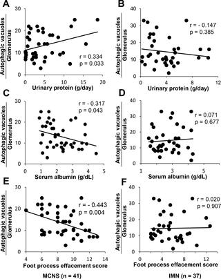
Autophagy is a cellular mechanism involved in the bulk degradation of proteins and turnover of organelle. Several studies have shown the significance of autophagy of the renal tubular epithelium in rodent models of tubulointerstitial disorder. However, the role of autophagy in the regulation of human glomerular diseases is largely unknown. The current study aimed to demonstrate morphological evidence of autophagy and its association with the ultrastructural changes of podocytes and clinical data in patients with idiopathic nephrotic syndrome, a disease in which patients exhibit podocyte injury. The study population included 95 patients, including patients with glomerular disease (minimal change nephrotic syndrome [MCNS], n = 41; idiopathic membranous nephropathy [IMN], n = 37) and 17 control subjects who underwent percutaneous renal biopsy. The number of autophagic vacuoles and the grade of foot process effacement (FPE) in podocytes were examined by electron microscopy (EM). The relationships among the expression of autophagic vacuoles, the grade of FPE, and the clinical data were determined. Autophagic vacuoles were mainly detected in podocytes by EM. The microtubule-associated protein 1 light chain 3 (LC3)-positive area was co-localized with the Wilms tumor 1 (WT1)-positive area on immunofluorescence microscopy, which suggested that autophagy occurred in the podocytes of patients with MCNS. The number of autophagic vacuoles in the podocytes was significantly correlated with the podocyte FPE score (r = -0.443, p = 0.004), the amount of proteinuria (r = 0.334, p = 0.033), and the level of serum albumin (r = -0.317, p = 0.043) in patients with MCNS. The FPE score was a significant determinant for autophagy after adjusting for the age in a multiple regression analysis in MCNS patients (p = 0.0456). However, such correlations were not observed in patients with IMN or in control subjects. In conclusion, the results indicated that the autophagy of podocytes is associated with FPE and severe proteinuria in patients with MCNS. The mechanisms underlying the activation of autophagy in association with FPE in podocytes should be further investigated in order to elucidate the pathophysiology of MCNS.
中文翻译:

肾小球改变综合征患者足细胞自噬与足突消失和蛋白尿有关。
自噬是一种参与蛋白质大量降解和细胞器更新的细胞机制。多项研究表明,在肾小管间质疾病的啮齿动物模型中,肾小管上皮自噬的重要性。然而,自噬在调节人类肾小球疾病中的作用尚不清楚。本研究旨在证明特发性肾病综合征患者自足吞噬的形态学证据及其与足细胞超微结构变化的关系以及临床数据,该病患者表现为足细胞损伤。研究人群包括95例患者,包括肾小球疾病患者(最小变化肾病综合征[MCNS],n = 41;特发性膜性肾病[IMN],n = 37)和17例接受了经皮肾穿刺活检的对照组。通过电子显微镜(EM)检查足细胞中自噬泡的数量和足突消失的程度(FPE)。确定了自噬泡的表达,FPE等级和临床数据之间的关系。自噬泡主要通过EM在足细胞中检测到。在免疫荧光显微镜下,微管相关蛋白1轻链3(LC3)阳性区域与Wilms肿瘤1(WT1)阳性区域共定位,这提示MCNS患者的足细胞发生自噬。足细胞自噬泡的数量与足细胞FPE评分(r = -0.443,p = 0.004),蛋白尿量(r = 0.334,p = 0.033)和血清白蛋白水平(r = -0.317,p = 0.043)。在MCNS患者的多元回归分析中,调整年龄后,FPE评分是自噬的重要决定因素(p = 0.0456)。然而,在IMN患者或对照受试者中未观察到这种相关性。总之,结果表明,MCNS患者足细胞的自噬与FPE和严重蛋白尿有关。为了阐明MCNS的病理生理,应进一步研究与足细胞中FPE结合的自噬激活的机制。结果表明,MCNS患者足细胞自噬与FPE和严重蛋白尿有关。为了阐明MCNS的病理生理,应进一步研究与足细胞中FPE结合的自噬激活的机制。结果表明,MCNS患者足细胞自噬与FPE和严重蛋白尿有关。为了阐明MCNS的病理生理,应进一步研究与足细胞中FPE结合的自噬激活的机制。
更新日期:2020-01-26
中文翻译:
肾小球改变综合征患者足细胞自噬与足突消失和蛋白尿有关。
自噬是一种参与蛋白质大量降解和细胞器更新的细胞机制。多项研究表明,在肾小管间质疾病的啮齿动物模型中,肾小管上皮自噬的重要性。然而,自噬在调节人类肾小球疾病中的作用尚不清楚。本研究旨在证明特发性肾病综合征患者自足吞噬的形态学证据及其与足细胞超微结构变化的关系以及临床数据,该病患者表现为足细胞损伤。研究人群包括95例患者,包括肾小球疾病患者(最小变化肾病综合征[MCNS],n = 41;特发性膜性肾病[IMN],n = 37)和17例接受了经皮肾穿刺活检的对照组。通过电子显微镜(EM)检查足细胞中自噬泡的数量和足突消失的程度(FPE)。确定了自噬泡的表达,FPE等级和临床数据之间的关系。自噬泡主要通过EM在足细胞中检测到。在免疫荧光显微镜下,微管相关蛋白1轻链3(LC3)阳性区域与Wilms肿瘤1(WT1)阳性区域共定位,这提示MCNS患者的足细胞发生自噬。足细胞自噬泡的数量与足细胞FPE评分(r = -0.443,p = 0.004),蛋白尿量(r = 0.334,p = 0.033)和血清白蛋白水平(r = -0.317,p = 0.043)。在MCNS患者的多元回归分析中,调整年龄后,FPE评分是自噬的重要决定因素(p = 0.0456)。然而,在IMN患者或对照受试者中未观察到这种相关性。总之,结果表明,MCNS患者足细胞的自噬与FPE和严重蛋白尿有关。为了阐明MCNS的病理生理,应进一步研究与足细胞中FPE结合的自噬激活的机制。结果表明,MCNS患者足细胞自噬与FPE和严重蛋白尿有关。为了阐明MCNS的病理生理,应进一步研究与足细胞中FPE结合的自噬激活的机制。结果表明,MCNS患者足细胞自噬与FPE和严重蛋白尿有关。为了阐明MCNS的病理生理,应进一步研究与足细胞中FPE结合的自噬激活的机制。


 京公网安备 11010802027423号