当前位置:
X-MOL 学术
›
Anal. Chim. Acta
›
论文详情
Our official English website, www.x-mol.net, welcomes your
feedback! (Note: you will need to create a separate account there.)
Effect of Sample Preparation Techniques upon Single Cell Chemical Imaging: A Practical Comparison between Synchrotron Radiation based X-ray Fluorescence (SR-XRF) and Nanoscopic Secondary Ion Mass Spectrometry (nano-SIMS)
Analytica Chimica Acta ( IF 5.7 ) Pub Date : 2020-04-01 , DOI: 10.1016/j.aca.2020.01.054 Björn De Samber , Riet De Rycke , Michiel De Bruyne , Michiel Kienhuis , Linda Sandblad , Sylvain Bohic , Peter Cloetens , Constantin Urban , Lubos Polerecky , Laszlo Vincze
Analytica Chimica Acta ( IF 5.7 ) Pub Date : 2020-04-01 , DOI: 10.1016/j.aca.2020.01.054 Björn De Samber , Riet De Rycke , Michiel De Bruyne , Michiel Kienhuis , Linda Sandblad , Sylvain Bohic , Peter Cloetens , Constantin Urban , Lubos Polerecky , Laszlo Vincze
|
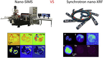
Analytical capabilities of Nanoscopic Secondary Ion Mass Spectrometry (nano-SIMS) and Synchrotron Radiation based X-ray Fluorescence (SR nano-XRF) techniques were compared for nanochemical imaging of polymorphonuclear human neutrophils (PMNs). PMNs were high pressure frozen (HPF), cryo-substituted, embedded in Spurr's resin and cut in thin sections (500 nm and 2 μm for both techniques resp.) Nano-SIMS enabled nanoscale mapping of isotopes of C, N, O, P and S, while SR based nano-XRF enabled trace level imaging of metals like Ca, Mn, Fe, Ni, Cu and Zn at a resolution of approx. 50 nm. The obtained elemental distributions were compared with those of whole, cryofrozen PMNs measured at the newly developed ID16A nano-imaging beamline at the European Synchrotron Radiation Facility (ESRF) in Grenoble, France. Similarities were observed for elements more tightly bound to the cell structure such as phosphorus and sulphur, while differences for mobile ions such as chlorine and potassium were more pronounced. Due to the observed elemental redistribution of mobile ions such as potassium and chlorine, elemental analysis of high pressure frozen (HPF), cryo-substituted and imbedded cells should be interpreted critically. Although decreasing analytical sensitivity occurs due to the presence of ice, analysis of cryofrozen cells - close to their native state - remains the golden standard. In general, we found nanoscale secondary ion mass spectrometry (nano-SIMS) and synchrotron radiation based nanoscopic X-ray fluorescence (SR nano-XRF) to be two supplementary alternatives for nanochemical imaging of single cells at the nanoscale.
中文翻译:

样品制备技术对单细胞化学成像的影响:基于同步辐射的 X 射线荧光 (SR-XRF) 与纳米二次离子质谱 (nano-SIMS) 的实际比较
比较了纳米级二次离子质谱 (nano-SIMS) 和基于同步辐射的 X 射线荧光 (SR nano-XRF) 技术的分析能力,用于多形核人中性粒细胞 (PMN) 的纳米化学成像。PMN 被高压冷冻 (HPF)、低温取代、嵌入 Spurr 的树脂中并切成薄片(两种技术分别为 500 nm 和 2 μm)纳米 SIMS 能够实现 C、N、O、P 同位素的纳米级映射和 S,而基于 SR 的纳米 XRF 能够以大约 100 的分辨率对 Ca、Mn、Fe、Ni、Cu 和 Zn 等金属进行痕量级成像。50 纳米。将获得的元素分布与在法国格勒诺布尔的欧洲同步加速器辐射设施 (ESRF) 新开发的 ID16A 纳米成像光束线测量的整个低温冷冻 PMN 的分布进行比较。观察到与细胞结构更紧密结合的元素(如磷和硫)的相似性,而移动离子(如氯和钾)的差异更为明显。由于观察到钾和氯等移动离子的元素重新分布,高压冷冻 (HPF)、低温置换和嵌入细胞的元素分析应严格解释。尽管由于冰的存在会降低分析灵敏度,但对冷冻细胞的分析 - 接近其天然状态 - 仍然是黄金标准。总的来说,我们发现纳米级二次离子质谱法 (nano-SIMS) 和基于同步辐射的纳米级 X 射线荧光 (SR nano-XRF) 是纳米级单细胞纳米化学成像的两种补充替代方法。
更新日期:2020-04-01
中文翻译:
样品制备技术对单细胞化学成像的影响:基于同步辐射的 X 射线荧光 (SR-XRF) 与纳米二次离子质谱 (nano-SIMS) 的实际比较
比较了纳米级二次离子质谱 (nano-SIMS) 和基于同步辐射的 X 射线荧光 (SR nano-XRF) 技术的分析能力,用于多形核人中性粒细胞 (PMN) 的纳米化学成像。PMN 被高压冷冻 (HPF)、低温取代、嵌入 Spurr 的树脂中并切成薄片(两种技术分别为 500 nm 和 2 μm)纳米 SIMS 能够实现 C、N、O、P 同位素的纳米级映射和 S,而基于 SR 的纳米 XRF 能够以大约 100 的分辨率对 Ca、Mn、Fe、Ni、Cu 和 Zn 等金属进行痕量级成像。50 纳米。将获得的元素分布与在法国格勒诺布尔的欧洲同步加速器辐射设施 (ESRF) 新开发的 ID16A 纳米成像光束线测量的整个低温冷冻 PMN 的分布进行比较。观察到与细胞结构更紧密结合的元素(如磷和硫)的相似性,而移动离子(如氯和钾)的差异更为明显。由于观察到钾和氯等移动离子的元素重新分布,高压冷冻 (HPF)、低温置换和嵌入细胞的元素分析应严格解释。尽管由于冰的存在会降低分析灵敏度,但对冷冻细胞的分析 - 接近其天然状态 - 仍然是黄金标准。总的来说,我们发现纳米级二次离子质谱法 (nano-SIMS) 和基于同步辐射的纳米级 X 射线荧光 (SR nano-XRF) 是纳米级单细胞纳米化学成像的两种补充替代方法。


 京公网安备 11010802027423号