当前位置:
X-MOL 学术
›
Aging Cell
›
论文详情
Our official English website, www.x-mol.net, welcomes your
feedback! (Note: you will need to create a separate account there.)
Tissue specificity of senescent cell accumulation during physiologic and accelerated aging of mice.
Aging Cell ( IF 8.0 ) Pub Date : 2020-01-25 , DOI: 10.1111/acel.13094 Matthew J Yousefzadeh 1, 2, 3 , Jing Zhao 3 , Christina Bukata 3, 4 , Erin A Wade 3, 4 , Sara J McGowan 1, 2, 3 , Luise A Angelini 1, 2, 3 , Michael P Bank 3, 5 , Aditi U Gurkar 3, 6 , Collin A McGuckian 1, 2, 3 , Mariah F Calubag 3, 4 , Jonathan I Kato 3, 4 , Christin E Burd 7 , Paul D Robbins 1, 2, 3 , Laura J Niedernhofer 1, 2, 3
Aging Cell ( IF 8.0 ) Pub Date : 2020-01-25 , DOI: 10.1111/acel.13094 Matthew J Yousefzadeh 1, 2, 3 , Jing Zhao 3 , Christina Bukata 3, 4 , Erin A Wade 3, 4 , Sara J McGowan 1, 2, 3 , Luise A Angelini 1, 2, 3 , Michael P Bank 3, 5 , Aditi U Gurkar 3, 6 , Collin A McGuckian 1, 2, 3 , Mariah F Calubag 3, 4 , Jonathan I Kato 3, 4 , Christin E Burd 7 , Paul D Robbins 1, 2, 3 , Laura J Niedernhofer 1, 2, 3
Affiliation
|
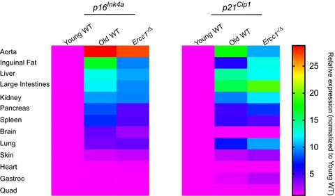
Senescent cells accumulate with age in vertebrates and promote aging largely through their senescence‐associated secretory phenotype (SASP). Many types of stress induce senescence, including genotoxic stress. ERCC1‐XPF is a DNA repair endonuclease required for multiple DNA repair mechanisms that protect the nuclear genome. Humans or mice with reduced expression of this enzyme age rapidly due to increased levels of spontaneous, genotoxic stress. Here, we asked whether this corresponds to an increased level of senescent cells. p16Ink4a and p21Cip1 mRNA were increased ~15‐fold in peripheral lymphocytes from 4‐ to 5‐month‐old Ercc1−/∆ and 2.5‐year‐old wild‐type (WT) mice, suggesting that these animals exhibit a similar biological age. p16Ink4a and p21Cip1 mRNA were elevated in 10 of 13 tissues analyzed from 4‐ to 5‐month‐old Ercc1−/∆ mice, indicating where endogenous DNA damage drives senescence in vivo. Aged WT mice had similar increases of p16Ink4a and p21Cip1 mRNA in the same 10 tissues as the mutant mice. Senescence‐associated β–galactosidase activity and p21Cip1 protein also were increased in tissues of the progeroid and aged mice, while Lamin B1 mRNA and protein levels were diminished. In Ercc1−/Δ mice with a p16Ink4a luciferase reporter, bioluminescence rose steadily with age, particularly in lung, thymus, and pancreas. These data illustrate where senescence occurs with natural and accelerated aging in mice and the relative extent of senescence among tissues. Interestingly, senescence was greater in male mice until the end of life. The similarities between Ercc1−/∆ and aged WT mice support the conclusion that the DNA repair‐deficient mice accurately model the age‐related accumulation of senescent cells, albeit six‐times faster.
中文翻译:

小鼠生理和加速衰老过程中衰老细胞积累的组织特异性。
衰老细胞在脊椎动物中随着年龄的增长而积累,并在很大程度上通过衰老相关的分泌表型(SASP)促进衰老。许多类型的压力会导致衰老,包括遗传毒性压力。ERCC1-XPF是一种DNA修复核酸内切酶,是保护核基因组的多种DNA修复机制所必需的。由于自发的遗传毒性应激水平升高,人类或小鼠的这种酶表达降低。在这里,我们问这是否对应于衰老细胞水平的增加。从4个月到5个月大的Ercc1- / ∆外周血中p16 Ink4a和p21 Cip1 mRNA增加了约15倍和2.5岁野生型(WT)小鼠,表明这些动物的生物学年龄相似。在4个月至5个月大的Ercc1- / ∆小鼠中,分析的13个组织中有10个组织中的10个组织中p16 Ink4a和p21 Cip1 mRNA升高,表明内源性DNA损伤在体内驱动衰老。老年WT小鼠在与突变小鼠相同的10个组织中p16 Ink4a和p21 Cip1 mRNA的增加相似。在衰老和衰老小鼠的组织中,衰老相关的β-半乳糖苷酶活性和p21 Cip1蛋白也增加,而Lamin B1 mRNA和蛋白水平降低。在Ercc1中-/Δ使用p16 Ink4a荧光素酶报道基因的小鼠,生物发光随年龄增长而稳定增长,尤其是在肺,胸腺和胰腺中。这些数据说明了在小鼠中自然衰老和加速衰老会发生衰老的地方,以及组织之间衰老的相对程度。有趣的是,直到寿命终结,雄性小鼠的衰老才更大。Ercc1- / ∆与衰老的WT小鼠之间的相似性支持以下结论:DNA修复缺陷小鼠可以准确地模拟衰老细胞与年龄相关的积累,尽管速度要快六倍。
更新日期:2020-01-25
中文翻译:
小鼠生理和加速衰老过程中衰老细胞积累的组织特异性。
衰老细胞在脊椎动物中随着年龄的增长而积累,并在很大程度上通过衰老相关的分泌表型(SASP)促进衰老。许多类型的压力会导致衰老,包括遗传毒性压力。ERCC1-XPF是一种DNA修复核酸内切酶,是保护核基因组的多种DNA修复机制所必需的。由于自发的遗传毒性应激水平升高,人类或小鼠的这种酶表达降低。在这里,我们问这是否对应于衰老细胞水平的增加。从4个月到5个月大的Ercc1- / ∆外周血中p16 Ink4a和p21 Cip1 mRNA增加了约15倍和2.5岁野生型(WT)小鼠,表明这些动物的生物学年龄相似。在4个月至5个月大的Ercc1- / ∆小鼠中,分析的13个组织中有10个组织中的10个组织中p16 Ink4a和p21 Cip1 mRNA升高,表明内源性DNA损伤在体内驱动衰老。老年WT小鼠在与突变小鼠相同的10个组织中p16 Ink4a和p21 Cip1 mRNA的增加相似。在衰老和衰老小鼠的组织中,衰老相关的β-半乳糖苷酶活性和p21 Cip1蛋白也增加,而Lamin B1 mRNA和蛋白水平降低。在Ercc1中-/Δ使用p16 Ink4a荧光素酶报道基因的小鼠,生物发光随年龄增长而稳定增长,尤其是在肺,胸腺和胰腺中。这些数据说明了在小鼠中自然衰老和加速衰老会发生衰老的地方,以及组织之间衰老的相对程度。有趣的是,直到寿命终结,雄性小鼠的衰老才更大。Ercc1- / ∆与衰老的WT小鼠之间的相似性支持以下结论:DNA修复缺陷小鼠可以准确地模拟衰老细胞与年龄相关的积累,尽管速度要快六倍。


 京公网安备 11010802027423号