当前位置:
X-MOL 学术
›
NMR Biomed.
›
论文详情
Our official English website, www.x-mol.net, welcomes your
feedback! (Note: you will need to create a separate account there.)
Quantification of regional myocardial mean intracellular water lifetime: A nonhuman primate study in myocardial stress.
NMR in Biomedicine ( IF 2.7 ) Pub Date : 2020-01-24 , DOI: 10.1002/nbm.4248 Smita Sampath 1 , Annamalai Sarayu Parimal 1 , Wei Huang 2 , Elaine Manigbas 3, 4 , Willy Gsell 3, 5 , Miko May Lee Chang 1 , Anqi Qiu 6 , Kirsten Jacobsen 7 , Jeffrey L Evelhoch 8 , Chih-Liang Chin 1
NMR in Biomedicine ( IF 2.7 ) Pub Date : 2020-01-24 , DOI: 10.1002/nbm.4248 Smita Sampath 1 , Annamalai Sarayu Parimal 1 , Wei Huang 2 , Elaine Manigbas 3, 4 , Willy Gsell 3, 5 , Miko May Lee Chang 1 , Anqi Qiu 6 , Kirsten Jacobsen 7 , Jeffrey L Evelhoch 8 , Chih-Liang Chin 1
Affiliation
|
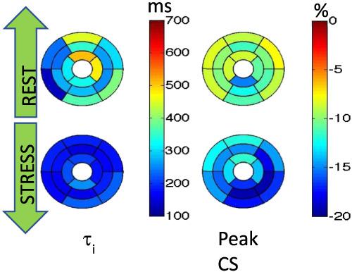
Heart failure with preserved ejection fraction (HFpEF) is typically associated with early metabolic remodeling. Noninvasive imaging biomarkers that reflect these changes will be crucial in determining responses to early drug interventions in these patients. Mean intracellular water lifetime (τi ) has been shown to be partially inversely related to Na, K-ATPase transporter activity and may thus provide insight into the metabolic status in HFpEF patients. Here, we aim to perform regional quantification of τi using dynamic contrast-enhanced (DCE) magnetic resonance imaging (MRI) in the nonhuman primate (NHP) heart and evaluate its region-specific variations under conditions of myocardial stress in the context of perturbed myocardial function. Cardiac stress was induced in seven naïve cynomolgus macaques using a dobutamine stepwise infusion protocol. All animals underwent 3 T cardiac dual-bolus DCE and tagging MRI experiments. The shutter-speed model was employed to quantify regional τi from the DCE-MR images. Additionally, τi values were correlated with myocardial strains. During cardiac stress, there was a significant decrease in global τi (192.9 ± 76.3 ms vs 321.6 ± 70 ms at rest, P < 0.05) in the left ventricle, together with an increase in global peak circumferential strain (-15.4% ± 2.7% vs -10.1% ± 2.9% at rest, P < 0.05). Specifically, slice-level analysis further revealed that a greater significant decrease in mean τi was observed in the apical region (ΔτI = 182.4 ms) compared with the basal (Δτi = 113.2 ms) and midventricular regions (Δτi = 108.4 ms). Regional analysis revealed that there was a greater significant decrease in mean τi in the anterior (Δτi = 243.9 ms) and antero-lateral (Δτi = 177.2 ms) regions. In the inferior and infero-septal regions, although a decrease in τi was observed, it was not significant. Whole heart regional quantification of τi is feasible using DCE-MRI. τi is sensitive to regional changes in metabolic state during cardiac stress, and its value correlates with strain.
中文翻译:

定量区域平均心肌细胞内水的寿命:一项关于心肌应激的非人类灵长类动物研究。
保留射血分数(HFpEF)的心力衰竭通常与早期代谢重构有关。反映这些变化的非侵入性成像生物标志物对于确定这些患者对早期药物干预的反应至关重要。已显示平均细胞内水寿命(τi)与Na,K-ATPase转运蛋白活性部分成反比,因此可以提供对HFpEF患者代谢状态的了解。在这里,我们旨在使用动态对比增强(DCE)磁共振成像(MRI)在非人类灵长类动物(NHP)心脏中对τi进行区域量化,并在心肌紧张的情况下评估心肌应激条件下其区域特异性变化功能。使用多巴酚丁胺逐步输注方案在七只幼稚食蟹猕猴中诱发心脏应激。所有动物均进行3 T心脏双剂量DCE和标记MRI实验。快门速度模型被用来从DCE-MR图像中量化区域τi。另外,τi值与心肌张力相关。在心脏应激期间,左心室的整体τi显着降低(192.9±76.3 ms vs静止时的321.6±70 ms,P <0.05),并且整体峰值周向应变增加(-15.4%±2.7%)与静止时的-10.1%±2.9%相比,P <0.05)。具体而言,切片水平分析进一步显示,与基础区域(Δτi= 113.2 ms)和心室中部区域(Δτi= 108.4 ms)相比,在顶端区域(ΔτI= 182.4 ms)观察到平均τi的更大降低。区域分析显示,前侧的平均τi显着更大下降(Δτi= 243。9毫秒)和前外侧(Δτi= 177.2毫秒)区域。在下部和中隔区,尽管观察到τi减小,但并不明显。使用DCE-MRI对τi进行全心区域量化是可行的。τi对心脏压力期间代谢状态的区域变化敏感,其值与应变相关。
更新日期:2020-03-09
中文翻译:
定量区域平均心肌细胞内水的寿命:一项关于心肌应激的非人类灵长类动物研究。
保留射血分数(HFpEF)的心力衰竭通常与早期代谢重构有关。反映这些变化的非侵入性成像生物标志物对于确定这些患者对早期药物干预的反应至关重要。已显示平均细胞内水寿命(τi)与Na,K-ATPase转运蛋白活性部分成反比,因此可以提供对HFpEF患者代谢状态的了解。在这里,我们旨在使用动态对比增强(DCE)磁共振成像(MRI)在非人类灵长类动物(NHP)心脏中对τi进行区域量化,并在心肌紧张的情况下评估心肌应激条件下其区域特异性变化功能。使用多巴酚丁胺逐步输注方案在七只幼稚食蟹猕猴中诱发心脏应激。所有动物均进行3 T心脏双剂量DCE和标记MRI实验。快门速度模型被用来从DCE-MR图像中量化区域τi。另外,τi值与心肌张力相关。在心脏应激期间,左心室的整体τi显着降低(192.9±76.3 ms vs静止时的321.6±70 ms,P <0.05),并且整体峰值周向应变增加(-15.4%±2.7%)与静止时的-10.1%±2.9%相比,P <0.05)。具体而言,切片水平分析进一步显示,与基础区域(Δτi= 113.2 ms)和心室中部区域(Δτi= 108.4 ms)相比,在顶端区域(ΔτI= 182.4 ms)观察到平均τi的更大降低。区域分析显示,前侧的平均τi显着更大下降(Δτi= 243。9毫秒)和前外侧(Δτi= 177.2毫秒)区域。在下部和中隔区,尽管观察到τi减小,但并不明显。使用DCE-MRI对τi进行全心区域量化是可行的。τi对心脏压力期间代谢状态的区域变化敏感,其值与应变相关。


 京公网安备 11010802027423号