当前位置:
X-MOL 学术
›
Nat. Protoc.
›
论文详情
Our official English website, www.x-mol.net, welcomes your
feedback! (Note: you will need to create a separate account there.)
Direct and simultaneous observation of transcription and chromosome architecture in single cells with Hi-M.
Nature Protocols ( IF 13.1 ) Pub Date : 2020-01-22 , DOI: 10.1038/s41596-019-0269-9 Andrés M Cardozo Gizzi 1, 2 , Sergio M Espinola 1 , Julian Gurgo 1 , Christophe Houbron 1 , Jean-Bernard Fiche 1 , Diego I Cattoni 1 , Marcelo Nollmann 1
Nature Protocols ( IF 13.1 ) Pub Date : 2020-01-22 , DOI: 10.1038/s41596-019-0269-9 Andrés M Cardozo Gizzi 1, 2 , Sergio M Espinola 1 , Julian Gurgo 1 , Christophe Houbron 1 , Jean-Bernard Fiche 1 , Diego I Cattoni 1 , Marcelo Nollmann 1
Affiliation
|
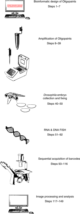
Simultaneous observation of 3D chromatin organization and transcription at the single-cell level and with high spatial resolution may hold the key to unveiling the mechanisms regulating embryonic development, cell differentiation and even disease. We recently developed Hi-M, a technology that enables the sequential labeling, 3D imaging and localization of multiple genomic DNA loci, together with RNA expression, in single cells within whole, intact Drosophila embryos. Importantly, Hi-M enables simultaneous detection of RNA expression and chromosome organization without requiring sample unmounting and primary probe rehybridization. Here, we provide a step-by-step protocol describing the design of probes, the preparation of samples, the stable immobilization of embryos in microfluidic chambers, and the complete procedure for image acquisition. The combined RNA/DNA fluorescence in situ hybridization procedure takes 4-5 d, including embryo collection. In addition, we describe image analysis software to segment nuclei, detect genomic spots, correct for drift and produce Hi-M matrices. A typical Hi-M experiment takes 1-2 d to complete all rounds of labeling and imaging and 4 additional days for image analysis. This technology can be easily expanded to investigate cell differentiation in cultured cells or organization of chromatin within complex tissues.
中文翻译:

直接和同时观察Hi-M在单个细胞中的转录和染色体结构。
在单细胞水平上以高空间分辨率同时观察3D染色质的组织和转录可能是揭示调节胚胎发育,细胞分化甚至疾病的机制的关键。我们最近开发了Hi-M,该技术可在完整完整的果蝇胚胎内的单个细胞中实现多个基因组DNA基因座的顺序标记,3D成像和定位以及RNA表达。重要的是,Hi-M能够同时检测RNA表达和染色体组织,而无需卸下样品和进行初级探针杂交。在这里,我们提供了一个分步协议,描述了探针的设计,样品的制备,将胚胎稳定地固定在微流体腔室内以及完整的图像采集程序。组合的RNA / DNA荧光原位杂交过程需要4-5 d,包括收集胚胎。此外,我们描述了图像分析软件以分割核,检测基因组斑点,校正漂移并产生Hi-M矩阵。一个典型的Hi-M实验需要1-2天才能完成所有的标记和成像过程,另外需要4天来进行图像分析。这项技术可以轻松扩展,以研究培养细胞的细胞分化或复杂组织内染色质的组织。一个典型的Hi-M实验需要1-2天才能完成所有的标记和成像过程,另外需要4天来进行图像分析。这项技术可以轻松扩展,以研究培养细胞的细胞分化或复杂组织内染色质的组织。一个典型的Hi-M实验需要1-2天才能完成所有的标记和成像过程,另外需要4天来进行图像分析。这项技术可以轻松扩展,以研究培养细胞的细胞分化或复杂组织内染色质的组织。
更新日期:2020-01-22
中文翻译:
直接和同时观察Hi-M在单个细胞中的转录和染色体结构。
在单细胞水平上以高空间分辨率同时观察3D染色质的组织和转录可能是揭示调节胚胎发育,细胞分化甚至疾病的机制的关键。我们最近开发了Hi-M,该技术可在完整完整的果蝇胚胎内的单个细胞中实现多个基因组DNA基因座的顺序标记,3D成像和定位以及RNA表达。重要的是,Hi-M能够同时检测RNA表达和染色体组织,而无需卸下样品和进行初级探针杂交。在这里,我们提供了一个分步协议,描述了探针的设计,样品的制备,将胚胎稳定地固定在微流体腔室内以及完整的图像采集程序。组合的RNA / DNA荧光原位杂交过程需要4-5 d,包括收集胚胎。此外,我们描述了图像分析软件以分割核,检测基因组斑点,校正漂移并产生Hi-M矩阵。一个典型的Hi-M实验需要1-2天才能完成所有的标记和成像过程,另外需要4天来进行图像分析。这项技术可以轻松扩展,以研究培养细胞的细胞分化或复杂组织内染色质的组织。一个典型的Hi-M实验需要1-2天才能完成所有的标记和成像过程,另外需要4天来进行图像分析。这项技术可以轻松扩展,以研究培养细胞的细胞分化或复杂组织内染色质的组织。一个典型的Hi-M实验需要1-2天才能完成所有的标记和成像过程,另外需要4天来进行图像分析。这项技术可以轻松扩展,以研究培养细胞的细胞分化或复杂组织内染色质的组织。


 京公网安备 11010802027423号