当前位置:
X-MOL 学术
›
NMR Biomed.
›
论文详情
Our official English website, www.x-mol.net, welcomes your
feedback! (Note: you will need to create a separate account there.)
Proton functional magnetic resonance spectroscopy in rodents
NMR in Biomedicine ( IF 2.7 ) Pub Date : 2020-01-22 , DOI: 10.1002/nbm.4254 Nathalie Just 1, 2
NMR in Biomedicine ( IF 2.7 ) Pub Date : 2020-01-22 , DOI: 10.1002/nbm.4254 Nathalie Just 1, 2
Affiliation
|
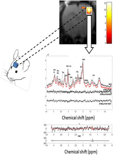
Proton functional magnetic resonance spectroscopy (1H‐fMRS) in the human brain is able to assess and quantify the metabolic response due to localized brain activity. Currently, 1H‐fMRS of the human brain is complementary to functional magnetic resonance imaging (fMRI) and a recommended technique at high field strengths (>7 T) for the investigation of neurometabolic couplings, thereby providing insight into the mechanisms underlying brain activity and brain connectivity. Understanding typical healthy brain metabolism during a task is expected to provide a baseline from which to detect and characterize neurochemical alterations associated with various neurological or psychiatric disorders and diseases. It is of paramount importance to resolve fundamental questions related to the regulation of neurometabolic processes. New techniques such as optogenetics may be coupled to fMRI and fMRS to bring more specificity to investigations of brain cell populations during cerebral activation thus enabling a higher link to molecular changes and therapeutic advances. These rather novel techniques are mainly available for rodent applications and trigger renewed interest in animal fMRS. However, rodent fMRS remains fairly confidential due to its inherent low signal‐to‐noise ratio and its dependence on anesthesia. For instance, the accurate determination of metabolic concentration changes during stimulation requires robust knowledge of the physiological environment of the measured region of interest linked to anesthesia in most cases. These factors may also have a strong influence on B0 homogeneity. Therefore, a degree of calibration of the stimulus strength and duration may be needed for increased knowledge of the underpinnings of cerebral activity. Here, we propose an early review of the current status of 1H‐fMRS in rodents and summarize current difficulties and future perspectives.
中文翻译:

啮齿动物的质子功能磁共振波谱
人脑中的质子功能磁共振波谱 ( 1 H-fMRS) 能够评估和量化由于局部大脑活动引起的代谢反应。目前,1人脑的 H-fMRS 是功能性磁共振成像 (fMRI) 的补充,也是一种推荐的高场强 (>7 T) 技术,用于研究神经代谢耦合,从而深入了解大脑活动和大脑连接的潜在机制。预计在一项任务中了解典型的健康大脑代谢将为检测和表征与各种神经或精神疾病和疾病相关的神经化学变化提供一个基线。解决与神经代谢过程调节相关的基本问题至关重要。光遗传学等新技术可以与 fMRI 和 fMRS 相结合,为大脑激活期间脑细胞群的研究带来更多特异性,从而与分子变化和治疗进展建立更高的联系。这些相当新颖的技术主要可用于啮齿动物应用,并引发对动物 fMRS 的重新兴趣。然而,由于其固有的低信噪比和对麻醉的依赖性,啮齿动物 fMRS 仍然相当保密。例如,在大多数情况下,刺激期间代谢浓度变化的准确测定需要对与麻醉相关的测量感兴趣区域的生理环境有充分的了解。这些因素也可能对 B0 同质性有很大影响。所以,为了增加对大脑活动基础的了解,可能需要对刺激强度和持续时间进行一定程度的校准。在这里,我们建议对目前的状况进行早期审查1 H-fMRS 在啮齿动物中的作用,并总结了当前的困难和未来的前景。
更新日期:2020-01-22
中文翻译:
啮齿动物的质子功能磁共振波谱
人脑中的质子功能磁共振波谱 ( 1 H-fMRS) 能够评估和量化由于局部大脑活动引起的代谢反应。目前,1人脑的 H-fMRS 是功能性磁共振成像 (fMRI) 的补充,也是一种推荐的高场强 (>7 T) 技术,用于研究神经代谢耦合,从而深入了解大脑活动和大脑连接的潜在机制。预计在一项任务中了解典型的健康大脑代谢将为检测和表征与各种神经或精神疾病和疾病相关的神经化学变化提供一个基线。解决与神经代谢过程调节相关的基本问题至关重要。光遗传学等新技术可以与 fMRI 和 fMRS 相结合,为大脑激活期间脑细胞群的研究带来更多特异性,从而与分子变化和治疗进展建立更高的联系。这些相当新颖的技术主要可用于啮齿动物应用,并引发对动物 fMRS 的重新兴趣。然而,由于其固有的低信噪比和对麻醉的依赖性,啮齿动物 fMRS 仍然相当保密。例如,在大多数情况下,刺激期间代谢浓度变化的准确测定需要对与麻醉相关的测量感兴趣区域的生理环境有充分的了解。这些因素也可能对 B0 同质性有很大影响。所以,为了增加对大脑活动基础的了解,可能需要对刺激强度和持续时间进行一定程度的校准。在这里,我们建议对目前的状况进行早期审查1 H-fMRS 在啮齿动物中的作用,并总结了当前的困难和未来的前景。


 京公网安备 11010802027423号