Our official English website, www.x-mol.net, welcomes your
feedback! (Note: you will need to create a separate account there.)
L-Plastin deficiency produces increased trabecular bone due to attenuation of sealing ring formation and osteoclast dysfunction.
Bone Research ( IF 14.3 ) Pub Date : 2020-01-22 , DOI: 10.1038/s41413-019-0079-2 Meenakshi A Chellaiah 1 , Megan C Moorer 2 , Sunipa Majumdar 1 , Hanan Aljohani 1 , Sharon C Morley 3 , Vanessa Yingling 4 , Joseph P Stains 2
Bone Research ( IF 14.3 ) Pub Date : 2020-01-22 , DOI: 10.1038/s41413-019-0079-2 Meenakshi A Chellaiah 1 , Megan C Moorer 2 , Sunipa Majumdar 1 , Hanan Aljohani 1 , Sharon C Morley 3 , Vanessa Yingling 4 , Joseph P Stains 2
Affiliation
|
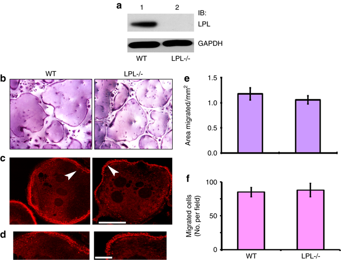
Bone resorption requires the formation of complex, actin-rich cytoskeletal structures. During the early phase of sealing ring formation by osteoclasts, L-plastin regulates actin-bundling to form the nascent sealing zones (NSZ). Here, we show that L-plastin knockout mice produce osteoclasts that are deficient in the formation of NSZs, are hyporesorptive, and make superficial resorption pits in vitro. Transduction of TAT-fused full-length L-plastin peptide into osteoclasts from L-plastin knockout mice rescued the formation of nascent sealing zones and sealing rings in a time-dependent manner. This response was not observed with mutated full-length L-plastin (Ser-5 and -7 to Ala-5 and -7) peptide. In contrast to the observed defect in the NSZ, L-plastin deficiency did not affect podosome formation or adhesion of osteoclasts in vitro or in vivo. Histomorphometry analyses in 8- and 12-week-old female L-plastin knockout mice demonstrated a decrease in eroded perimeters and an increase in trabecular bone density, without a change in bone formation by osteoblasts. This decrease in eroded perimeters supports that osteoclast function is attenuated in L-plastin knockouts. Micro-CT analyses confirmed a marked increase in trabecular bone mass. In conclusion, female L-plastin knockout mice had increased trabecular bone density due to impaired bone resorption by osteoclasts. L-plastin could be a potential target for therapeutic interventions to treat trabecular bone loss.
中文翻译:

L-塑蛋白缺乏会由于密封环形成减弱和破骨细胞功能障碍而导致骨小梁增加。
骨吸收需要形成复杂的、富含肌动蛋白的细胞骨架结构。在破骨细胞形成密封环的早期阶段,L-塑性蛋白调节肌动蛋白成束以形成新生密封区(NSZ)。在这里,我们发现 L-plastin 敲除小鼠产生的破骨细胞缺乏 NSZ 的形成,吸收性低下,并在体外形成浅表吸收凹坑。将 TAT 融合的全长 L-塑性蛋白肽转导至 L-塑性蛋白敲除小鼠的破骨细胞中,以时间依赖性方式挽救了新生密封区和密封环的形成。对于突变的全长 L-塑蛋白(Ser-5 和 -7 至 Ala-5 和 -7)肽,没有观察到这种反应。与观察到的 NSZ 缺陷相反,L-plastin 缺乏并不影响体外或体内破骨细胞足小体的形成或粘附。对 8 周和 12 周龄雌性 L-plastin 敲除小鼠进行的组织形态计量学分析表明,侵蚀周长减少,骨小梁密度增加,但成骨细胞的骨形成没有变化。侵蚀周长的减少支持了 L-塑性蛋白敲除中破骨细胞功能减弱。显微 CT 分析证实小梁骨量显着增加。总之,由于破骨细胞的骨吸收受损,雌性 L-plastin 敲除小鼠的骨小梁密度增加。 L-塑蛋白可能是治疗小梁骨丢失的治疗干预的潜在目标。
更新日期:2020-01-22
中文翻译:
L-塑蛋白缺乏会由于密封环形成减弱和破骨细胞功能障碍而导致骨小梁增加。
骨吸收需要形成复杂的、富含肌动蛋白的细胞骨架结构。在破骨细胞形成密封环的早期阶段,L-塑性蛋白调节肌动蛋白成束以形成新生密封区(NSZ)。在这里,我们发现 L-plastin 敲除小鼠产生的破骨细胞缺乏 NSZ 的形成,吸收性低下,并在体外形成浅表吸收凹坑。将 TAT 融合的全长 L-塑性蛋白肽转导至 L-塑性蛋白敲除小鼠的破骨细胞中,以时间依赖性方式挽救了新生密封区和密封环的形成。对于突变的全长 L-塑蛋白(Ser-5 和 -7 至 Ala-5 和 -7)肽,没有观察到这种反应。与观察到的 NSZ 缺陷相反,L-plastin 缺乏并不影响体外或体内破骨细胞足小体的形成或粘附。对 8 周和 12 周龄雌性 L-plastin 敲除小鼠进行的组织形态计量学分析表明,侵蚀周长减少,骨小梁密度增加,但成骨细胞的骨形成没有变化。侵蚀周长的减少支持了 L-塑性蛋白敲除中破骨细胞功能减弱。显微 CT 分析证实小梁骨量显着增加。总之,由于破骨细胞的骨吸收受损,雌性 L-plastin 敲除小鼠的骨小梁密度增加。 L-塑蛋白可能是治疗小梁骨丢失的治疗干预的潜在目标。


 京公网安备 11010802027423号