Our official English website, www.x-mol.net, welcomes your
feedback! (Note: you will need to create a separate account there.)
Intravital optoacoustic and ultrasound bio-microscopy reveal radiation-inhibited skull angiogenesis
Bone ( IF 3.5 ) Pub Date : 2020-04-01 , DOI: 10.1016/j.bone.2020.115251 Héctor Estrada 1 , Johannes Rebling 1 , Wolfgang Sievert 2 , Daniela Hladik 3 , Urs Hofmann 1 , Sven Gottschalk 4 , Soile Tapio 3 , Gabriele Multhoff 5 , Daniel Razansky 6
Bone ( IF 3.5 ) Pub Date : 2020-04-01 , DOI: 10.1016/j.bone.2020.115251 Héctor Estrada 1 , Johannes Rebling 1 , Wolfgang Sievert 2 , Daniela Hladik 3 , Urs Hofmann 1 , Sven Gottschalk 4 , Soile Tapio 3 , Gabriele Multhoff 5 , Daniel Razansky 6
Affiliation
|
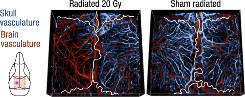
Angiogenesis is critical in bone development and growth. Dense, large-scale, and multi-layered vascular networks formed by thin-walled sinusoidal vessels perfuse the plate bones and play an important role in bone repair. Yet, the intricate functional morphology of skull microvasculature remains poorly understood as it is difficult to visualize using existing intravital microscopy techniques. Here we introduced an intravital fully-transcranial imaging approach based on hybrid optoacoustic and ultrasound bio-microscopy for large-scale observations and quantitative analysis of the vascular morphology, angiogenesis, vessel remodeling, and subsurface roughness in murine skulls. Our approach revealed radiation-inhibited angiogenesis in the skull bone. We also observed previously undocumented sinusoidal vascular networks spanning the entire skullcap, thus opening new vistas for studying the complex interactions between calvarial, pial, and cortical vascular systems.
中文翻译:

活体光声和超声生物显微镜揭示辐射抑制颅骨血管生成
血管生成对骨骼发育和生长至关重要。由薄壁正弦血管形成的致密、大范围、多层的血管网络灌注板骨,在骨修复中发挥重要作用。然而,颅骨微血管系统的复杂功能形态仍然知之甚少,因为使用现有的活体显微镜技术很难可视化。在这里,我们介绍了一种基于混合光声和超声生物显微镜的活体全经颅成像方法,用于对小鼠头骨中的血管形态、血管生成、血管重塑和表面粗糙度进行大规模观察和定量分析。我们的方法揭示了颅骨中辐射抑制的血管生成。我们还观察到了以前未记录的横跨整个头盖骨的正弦血管网络,
更新日期:2020-04-01
中文翻译:
活体光声和超声生物显微镜揭示辐射抑制颅骨血管生成
血管生成对骨骼发育和生长至关重要。由薄壁正弦血管形成的致密、大范围、多层的血管网络灌注板骨,在骨修复中发挥重要作用。然而,颅骨微血管系统的复杂功能形态仍然知之甚少,因为使用现有的活体显微镜技术很难可视化。在这里,我们介绍了一种基于混合光声和超声生物显微镜的活体全经颅成像方法,用于对小鼠头骨中的血管形态、血管生成、血管重塑和表面粗糙度进行大规模观察和定量分析。我们的方法揭示了颅骨中辐射抑制的血管生成。我们还观察到了以前未记录的横跨整个头盖骨的正弦血管网络,


 京公网安备 11010802027423号