Our official English website, www.x-mol.net, welcomes your
feedback! (Note: you will need to create a separate account there.)
18F-FDG-PET/MRI in the diagnostic work-up of limbic encephalitis.
PLOS ONE ( IF 2.9 ) Pub Date : 2020-01-17 , DOI: 10.1371/journal.pone.0227906 Cornelius Deuschl 1 , Theodor Rüber 2 , Leon Ernst 2 , Wolfgang P Fendler 3 , Julian Kirchner 4 , Christoph Mönninghoff 1, 5 , Ken Herrmann 3 , Carlos M Quesada 6 , Michael Forsting 1 , Christian E Elger 2 , Lale Umutlu 1
PLOS ONE ( IF 2.9 ) Pub Date : 2020-01-17 , DOI: 10.1371/journal.pone.0227906 Cornelius Deuschl 1 , Theodor Rüber 2 , Leon Ernst 2 , Wolfgang P Fendler 3 , Julian Kirchner 4 , Christoph Mönninghoff 1, 5 , Ken Herrmann 3 , Carlos M Quesada 6 , Michael Forsting 1 , Christian E Elger 2 , Lale Umutlu 1
Affiliation

|
INTRODUCTION
Limbic encephalitis (LE) is an immune-related, sometimes paraneoplastic process of the central nervous system. Initial diagnosis and treatment are based on the clinical presentation as well as antibody profiles and MRI. This study investigated the diagnostic value of integrated 18F-FDG-PET/MRI in the diagnostic work-up of patients with LE for a cerebral and whole-body imaging concept.
MATERIAL AND METHODS
Twenty patients with suspected LE were enrolled in this prospective study. All patients underwent a dedicated PET/MRI protocol of the brain as well as the whole-body. Two neuroradiologists, one body radiologist and one nuclear medicine physician performed blinded consensus readings of each corresponding MRI and PET/MRI dataset of the brain and whole-body. Diagnostic confidence was evaluated on a Likert scale.
RESULTS
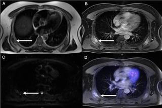
Based on integrated PET/MRI 19 / 20 patients were found to show morphologic and / or metabolic changes indicative of LE, whereas sole MRI enabled correct identification in 16 / 20 patients. Three patients with negative MRI showed metabolic changes of the limbic system or extra-limbic regions, shifting the diagnosis from (negative) MRI to positive for LE in PET/MRI. Whole-body staging revealed suspected lesions in 2/20 patients, identified by MRI and PET, one confirmed as malignant and one false positive. Diagnostic confidence for cerebral and whole-body imaging reached higher scores for PET/MRI (cerebral: 2.7 and whole body: 4.8) compared to MRI alone (cerebral: 2.4 and whole body: 4.5).
CONCLUSION
LE diagnosis remains challenging for imaging as it shows only subtle imaging findings in most patients. Nevertheless, based on the simultaneous and combined analysis of morphologic and metabolic data, integrated PET/MRI may enable a dual platform for improved diagnostic confidence and overall detection of LE as well as whole-body imaging for exclusion of paraneoplastic LE.
中文翻译:

18F-FDG-PET / MRI在边缘性脑炎的诊断中的作用。
简介边缘性脑炎(LE)是与免疫相关的,有时是中枢神经系统的副肿瘤过程。初步诊断和治疗基于临床表现以及抗体谱和MRI。这项研究调查了18F-FDG-PET / MRI集成在LE患者脑部和全身成像概念诊断中的诊断价值。材料与方法该前瞻性研究纳入了20名疑似LE的患者。所有患者均接受了大脑以及整个身体的专用PET / MRI方案。两名神经放射学家,一名身体放射学家和一名核医学医师对大脑和全身的每个相应MRI和PET / MRI数据集进行了盲目共有读数。诊断置信度用李克特量表评估。结果基于综合PET / MRI,发现19/20例患者表现出指示LE的形态和/或代谢变化,而单独的MRI能够正确鉴定16/20例患者。3例MRI阴性的患者显示出边缘系统或边缘周围区域的代谢变化,从而使PET / MRI诊断从(阴性)MRI变为阳性。全身分期显示在2/20例患者中有可疑病变,通过MRI和PET鉴别,其中1例被确认为恶性,1例为假阳性。与单独的MRI(大脑:2.4和全身:4.5)相比,PET / MRI(大脑:2.7和全身:4.8)对大脑和全身成像的诊断置信度更高。结论LE诊断对于影像学仍然具有挑战性,因为它在大多数患者中仅显示出细微的影像学发现。不过,
更新日期:2020-01-21
中文翻译:

18F-FDG-PET / MRI在边缘性脑炎的诊断中的作用。
简介边缘性脑炎(LE)是与免疫相关的,有时是中枢神经系统的副肿瘤过程。初步诊断和治疗基于临床表现以及抗体谱和MRI。这项研究调查了18F-FDG-PET / MRI集成在LE患者脑部和全身成像概念诊断中的诊断价值。材料与方法该前瞻性研究纳入了20名疑似LE的患者。所有患者均接受了大脑以及整个身体的专用PET / MRI方案。两名神经放射学家,一名身体放射学家和一名核医学医师对大脑和全身的每个相应MRI和PET / MRI数据集进行了盲目共有读数。诊断置信度用李克特量表评估。结果基于综合PET / MRI,发现19/20例患者表现出指示LE的形态和/或代谢变化,而单独的MRI能够正确鉴定16/20例患者。3例MRI阴性的患者显示出边缘系统或边缘周围区域的代谢变化,从而使PET / MRI诊断从(阴性)MRI变为阳性。全身分期显示在2/20例患者中有可疑病变,通过MRI和PET鉴别,其中1例被确认为恶性,1例为假阳性。与单独的MRI(大脑:2.4和全身:4.5)相比,PET / MRI(大脑:2.7和全身:4.8)对大脑和全身成像的诊断置信度更高。结论LE诊断对于影像学仍然具有挑战性,因为它在大多数患者中仅显示出细微的影像学发现。不过,


 京公网安备 11010802027423号