Our official English website, www.x-mol.net, welcomes your
feedback! (Note: you will need to create a separate account there.)
Pupil response to noxious corneal stimulation.
PLOS ONE ( IF 2.9 ) Pub Date : 2020-01-17 , DOI: 10.1371/journal.pone.0227771 Emmanuel B Alabi 1 , Trefford L Simpson 1
PLOS ONE ( IF 2.9 ) Pub Date : 2020-01-17 , DOI: 10.1371/journal.pone.0227771 Emmanuel B Alabi 1 , Trefford L Simpson 1
Affiliation
|
PURPOSE
Ocular somatosensory-autonomic reflexes play critical roles in maintaining homeostasis of the eye. The purpose of this study was to investigate the pupil response to nociceptive corneal stimuli.
METHODS
A Waterloo-Belmonte pneumatic esthesiometer was used to determine detection thresholds and randomly deliver mechanical and chemical stimuli from levels of detection threshold to twice the threshold in 50% steps to the central cornea of 15 healthy subjects. For each stimulus, imaging of the stimulated/unstimulated eye was performed using two modified/calibrated Logitech c920 digital cameras for 4 seconds each, pre/post stimulus capture. The data were processed with a custom segmentation algorithm to help identify the pupils and pupil diameter was measured using ImageJ software. Pupil dilation response differences between the ipsi- and contralateral eye was analyzed using dependent t-tests. The effect of stimulus intensity, modality and sex of subjects were analyzed using repeated measures.
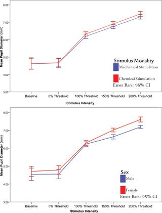
RESULTS
In mechanical and chemical stimulation experiments, there was no difference in pupil responses between the stimulated eye and the unstimulated eye, (all dependent T-test p > 0.05). On average, pupil diameter increased from baseline as the corneal stimulus intensity increased. This happened regardless of whether mechanical or chemical stimulation occurred (ANOVA p < 0.05). At 200% threshold, pupil diameter was greater than at all stimulus intensities (Tukey HSD, all p < 0.05). Based on stimulus intensity, females had greater pupil diameters than males at levels of 150% threshold and 200% threshold (ANOVA p < 0.05, all Tukey HSD p < 0.05).
CONCLUSION
This study serves as a basis for the characterization of the local stimulus-response neural circuitry relating nociceptive stimuli to autonomic responses and in combination with our work on completely separate autonomic circuits of bulbar conjunctival vessel dilation and reflex tearing suggests that the monotonic measurements of redness, tearing and pupils provide accurate, separable responses that reflect painful stimulus intensity.
中文翻译:

学生对有害角膜刺激的反应。
目的眼部的体感自主神经反射在维持眼内稳态方面起着至关重要的作用。这项研究的目的是调查瞳孔对伤害性角膜刺激的反应。方法使用滑铁卢-贝尔蒙特气动美学仪确定检测阈值,并将机械和化学刺激从检测阈值水平到阈值的两倍以50%的步长随机传递到15位健康受试者的中央角膜。对于每种刺激,使用两台经过修改/校准的Logitech c920数码相机对刺激/未刺激的眼睛进行成像,每次进行4秒钟,刺激之前/之后的捕捉。使用自定义分割算法处理数据以帮助识别瞳孔,并使用ImageJ软件测量瞳孔直径。使用相关的t检验分析同侧和对侧眼的瞳孔扩张反应差异。使用重复测量方法分析刺激强度,模态和性别对受试者的影响。结果在机械和化学刺激实验中,受刺激的眼睛和未受刺激的眼睛之间的瞳孔反应没有差异(所有相关的T检验p> 0.05)。平均而言,随着角膜刺激强度的增加,瞳孔直径从基线开始增加。无论是否发生机械或化学刺激,均会发生这种情况(ANOVA p <0.05)。在阈值200%时,瞳孔直径大于所有刺激强度(Tukey HSD,所有p <0.05)。根据刺激强度,在阈值150%和阈值200%时,女性的瞳孔直径大于男性(ANOVA p <0.05,所有Tukey HSD p <0.05)。结论这项研究为表征伤害感受性刺激与自主反应相关的局部刺激响应神经回路提供了基础,并结合我们对球结膜血管扩张和反射性撕裂的完全独立自主回路的研究表明,红色的单调测量,流泪和瞳孔提供准确,可分离的反应,反映出痛苦的刺激强度。
更新日期:2020-01-21
中文翻译:
学生对有害角膜刺激的反应。
目的眼部的体感自主神经反射在维持眼内稳态方面起着至关重要的作用。这项研究的目的是调查瞳孔对伤害性角膜刺激的反应。方法使用滑铁卢-贝尔蒙特气动美学仪确定检测阈值,并将机械和化学刺激从检测阈值水平到阈值的两倍以50%的步长随机传递到15位健康受试者的中央角膜。对于每种刺激,使用两台经过修改/校准的Logitech c920数码相机对刺激/未刺激的眼睛进行成像,每次进行4秒钟,刺激之前/之后的捕捉。使用自定义分割算法处理数据以帮助识别瞳孔,并使用ImageJ软件测量瞳孔直径。使用相关的t检验分析同侧和对侧眼的瞳孔扩张反应差异。使用重复测量方法分析刺激强度,模态和性别对受试者的影响。结果在机械和化学刺激实验中,受刺激的眼睛和未受刺激的眼睛之间的瞳孔反应没有差异(所有相关的T检验p> 0.05)。平均而言,随着角膜刺激强度的增加,瞳孔直径从基线开始增加。无论是否发生机械或化学刺激,均会发生这种情况(ANOVA p <0.05)。在阈值200%时,瞳孔直径大于所有刺激强度(Tukey HSD,所有p <0.05)。根据刺激强度,在阈值150%和阈值200%时,女性的瞳孔直径大于男性(ANOVA p <0.05,所有Tukey HSD p <0.05)。结论这项研究为表征伤害感受性刺激与自主反应相关的局部刺激响应神经回路提供了基础,并结合我们对球结膜血管扩张和反射性撕裂的完全独立自主回路的研究表明,红色的单调测量,流泪和瞳孔提供准确,可分离的反应,反映出痛苦的刺激强度。


 京公网安备 11010802027423号