Applied Materials Today ( IF 7.2 ) Pub Date : 2020-01-20 , DOI: 10.1016/j.apmt.2020.100561 Annie Agnes Suganya Samson , Sera Hong , Baskaran Purushothaman , Jeongmin Lee , Joon Myong Song
|
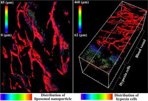
Passive targeting of nanoparticles (NPs) mediated by Enhanced Permeability and Retention effect is essential to NP delivery to tumors. However, clinically, only limited success has been found due to inadequate intratumoral delivery. Herein, for the first time, we present direct a demonstration visualizing the spatial distribution of liposomal nanoparticles (NP1: Lip-Ru (∼126 nm), NP2: Lip-Ru (∼164 nm) and NP3: Lip-Ru (∼234 nm)) in relation to hypoxic (HIF1α) and clonogenic (CD44) cells in intact transparent tumor tissues. Quantitative verification indicated that NP1, NP2, and NP3 did not diffuse into the intratumoral region over 100 μm away from the blood vessel. However, the liposomal nanoparticles represented slight difference in the three dimensional distribution within 100 μm around the blood vessel. Spatial distribution of HIF1α cells in tumors ranged from 62 to ∼460 μm. By contrast, CD44 expressing cells were observed in regions adjacent and away from the vessels. However, the maximum distribution of CD44 was observed ∼300 μm from the nearest vessels. Thus, the transparent tumor model reveals the penetration limits of Lip-Ru, and beyond the maximum diffusion of Lip-Ru, tumors contain hypoxic and clonogenic cells that are viable.
中文翻译:
透明的肿瘤微环境:脂质体纳米颗粒是否足以将药物递送至缺氧区域和克隆细胞?
由增强的渗透性和保留作用介导的纳米粒子(NPs)的被动靶向对于NP传递至肿瘤至关重要。然而,由于肿瘤内递送不足,在临床上仅发现有限的成功。在本文中,我们首次直接展示了脂质体纳米颗粒(NP1:Lip-Ru(〜126 nm),NP2:Lip-Ru(〜164 nm)和NP3:Lip-Ru(〜234)的空间分布完整透明肿瘤组织中的低氧(HIF1α)和克隆形成(CD44)细胞。定量验证表明,NP1,NP2和NP3不会扩散到距血管100μm的肿瘤内区域。然而,脂质体纳米颗粒在血管周围100μm内的三维分布中表现出微小的差异。HIF1α细胞在肿瘤中的空间分布范围为62至460μm。相反,在邻近和远离血管的区域中观察到表达CD44的细胞。但是,从最近的血管中观察到CD44的最大分布约为300μm。因此,透明的肿瘤模型揭示了Lip-Ru的渗透极限,并且除了Lip-Ru的最大扩散以外,肿瘤还包含低氧和克隆细胞。


 京公网安备 11010802027423号