当前位置:
X-MOL 学术
›
ACS Biomater. Sci. Eng.
›
论文详情
Our official English website, www.x-mol.net, welcomes your
feedback! (Note: you will need to create a separate account there.)
TiO2-Based Nanotopographical Cues Attenuate the Restenotic Phenotype in Primary Human Vascular Endothelial and Smooth Muscle Cells.
ACS Biomaterials Science & Engineering ( IF 5.4 ) Pub Date : 2020-01-17 , DOI: 10.1021/acsbiomaterials.9b01475 Yiqi Cao 1 , Tejal A Desai 2
ACS Biomaterials Science & Engineering ( IF 5.4 ) Pub Date : 2020-01-17 , DOI: 10.1021/acsbiomaterials.9b01475 Yiqi Cao 1 , Tejal A Desai 2
Affiliation

|
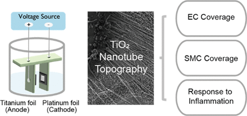
Coronary and peripheral stents are implants that are inserted into blocked arteries to restore blood flow. After stent deployment, the denudation of the endothelial cell (EC) layer and the resulting inflammatory cascade can lead to restenosis, the renarrowing of the vessel wall due to the hyperproliferation and excessive matrix secretion of smooth muscle cells (SMCs). Despite advances in drug-eluting stents (DES), restenosis remains a clinical challenge and can require repeat revascularizations. In this study, we investigated how vascular cell phenotype can be modulated by nanotopographical cues on the stent surface, with the goal of developing an alternative strategy to DES for decreasing restenosis. We fabricated TiO2 nanotubes and demonstrated that this topography can decrease SMC surface coverage without affecting endothelialization. In addition, to our knowledge, this is the first study reporting that TiO2 nanotube topography dampens the response to inflammatory cytokine stimulation in both endothelial and smooth muscle cells. We observed that compared to flat titanium surfaces, nanotube surfaces attenuated tumor necrosis factor alpha (TNFα)-induced vascular cell adhesion molecule-1 (VCAM-1) expression in ECs by 1.8-fold and decreased TNFα-induced SMC growth by 42%. Further, we found that the resulting cellular phenotype is sensitive to changes in nanotube diameter and that 90 nm diameter nanotubes leads to the greatest magnitude in cell response compared to 30 or 50 nm nanotubes.
中文翻译:

基于 TiO2 的纳米拓扑信号可减弱原代人血管内皮细胞和平滑肌细胞的再狭窄表型。
冠状动脉和外周支架是插入阻塞动脉以恢复血流的植入物。支架展开后,内皮细胞(EC)层的剥脱以及由此产生的炎症级联反应可导致再狭窄,由于平滑肌细胞(SMC)的过度增殖和过度基质分泌而导致血管壁重新狭窄。尽管药物洗脱支架 (DES) 取得了进展,但再狭窄仍然是一个临床挑战,可能需要重复血运重建。在这项研究中,我们研究了如何通过支架表面的纳米拓扑信号来调节血管细胞表型,目的是开发一种 DES 的替代策略来减少再狭窄。我们制造了 TiO 2纳米管,并证明这种形貌可以减少 SMC 表面覆盖而不影响内皮化。此外,据我们所知,这是第一项报告 TiO 2纳米管形貌抑制内皮细胞和平滑肌细胞对炎症细胞因子刺激的反应的研究。我们观察到,与平面钛表面相比,纳米管表面使 EC 中肿瘤坏死因子 α (TNFα) 诱导的血管细胞粘附分子 1 (VCAM-1) 表达减弱 1.8 倍,并使 TNFα 诱导的 SMC 生长降低 42%。此外,我们发现所得的细胞表型对纳米管直径的变化敏感,并且与 30 或 50 nm 纳米管相比,90 nm 直径的纳米管导致最大幅度的细胞反应。
更新日期:2020-01-17
中文翻译:

基于 TiO2 的纳米拓扑信号可减弱原代人血管内皮细胞和平滑肌细胞的再狭窄表型。
冠状动脉和外周支架是插入阻塞动脉以恢复血流的植入物。支架展开后,内皮细胞(EC)层的剥脱以及由此产生的炎症级联反应可导致再狭窄,由于平滑肌细胞(SMC)的过度增殖和过度基质分泌而导致血管壁重新狭窄。尽管药物洗脱支架 (DES) 取得了进展,但再狭窄仍然是一个临床挑战,可能需要重复血运重建。在这项研究中,我们研究了如何通过支架表面的纳米拓扑信号来调节血管细胞表型,目的是开发一种 DES 的替代策略来减少再狭窄。我们制造了 TiO 2纳米管,并证明这种形貌可以减少 SMC 表面覆盖而不影响内皮化。此外,据我们所知,这是第一项报告 TiO 2纳米管形貌抑制内皮细胞和平滑肌细胞对炎症细胞因子刺激的反应的研究。我们观察到,与平面钛表面相比,纳米管表面使 EC 中肿瘤坏死因子 α (TNFα) 诱导的血管细胞粘附分子 1 (VCAM-1) 表达减弱 1.8 倍,并使 TNFα 诱导的 SMC 生长降低 42%。此外,我们发现所得的细胞表型对纳米管直径的变化敏感,并且与 30 或 50 nm 纳米管相比,90 nm 直径的纳米管导致最大幅度的细胞反应。


 京公网安备 11010802027423号