Our official English website, www.x-mol.net, welcomes your
feedback! (Note: you will need to create a separate account there.)
Measurement of Prostate Volume with MRI (A Guide for the Perplexed): Biproximate Method with Analysis of Precision and Accuracy.
Scientific Reports ( IF 3.8 ) Pub Date : 2020-01-17 , DOI: 10.1038/s41598-019-57046-x Neil F Wasserman 1 , Eric Niendorf 1 , Benjamin Spilseth 1
Scientific Reports ( IF 3.8 ) Pub Date : 2020-01-17 , DOI: 10.1038/s41598-019-57046-x Neil F Wasserman 1 , Eric Niendorf 1 , Benjamin Spilseth 1
Affiliation
|
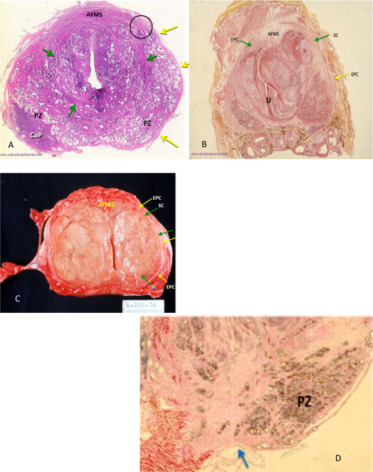
To review the anatomic basis of prostate boundary selection on T2-weighted magnetic resonance imaging (MRI). To introduce an alternative 3D ellipsoid measuring technique that maximizes precision, report the intra- and inter-observer reliability, and to advocate it's use for research involving multiple observers. We demonstrate prostate boundary anatomy using gross pathology and MRI examples. This provides background for selecting key boundary marks when measuring prostate volume. An alternative ellipsoid volume method is then proposed using these boundaries in an attempt to improve inter-observer precision. An IRB approved retrospective study of 140 patients with elevated serum prostate specific antigen levels and/or abnormal digital rectal examinations was done with T2-weighted MRI applying a new (Biproximate) technique. Measurements were made by 2 examiners, correlated with each other for inter-observer precision and correlated with an expert observer for accuracy. Correlation statistics, linear regression analysis, and tests of means were applied using p ≤ 0.05 as the threshold for significance. Inter-observer correlation (precision) was 0.95 between observers. Correlation between these observers and the expert (accuracy) was 0.94 and 0.97 respectively. Intra-observer correlation for the expert was 0.98. Means for inter-rater reliability and accuracy were all the same (p = 0.001). We conclude that using more precise reproducible landmarks with biproximate technique, precision and accuracy of total prostate volume is found to be demonstrated.
中文翻译:

用 MRI 测量前列腺体积(困惑者指南):双近似方法及精度和准确度分析。
回顾 T2 加权磁共振成像 (MRI) 上前列腺边界选择的解剖学基础。介绍一种可最大限度提高精度的替代 3D 椭球测量技术,报告观察者内和观察者间的可靠性,并提倡将其用于涉及多个观察者的研究。我们使用大体病理学和 MRI 示例展示前列腺边界解剖结构。这为测量前列腺体积时选择关键边界标记提供了背景。然后,使用这些边界提出了另一种椭球体体积方法,试图提高观察者间的精度。 IRB 批准了一项回顾性研究,对 140 名血清前列腺特异性抗原水平升高和/或直肠指检异常的患者进行了回顾性研究,该研究采用 T2 加权 MRI 应用新的(Biproximate)技术进行。测量由 2 名检查员进行,相互关联以确保观察者间的精确度,并与专家观察者关联以确保准确性。使用 p ≤ 0.05 作为显着性阈值,应用相关统计、线性回归分析和均值检验。观察者之间的观察者间相关性(精度)为 0.95。这些观察者和专家之间的相关性(准确度)分别为 0.94 和 0.97。专家的观察者内相关性为 0.98。评估者间的可靠性和准确性均相同 (p = 0.001)。我们的结论是,使用双近似技术的更精确的可重复性标志,可以证明总前列腺体积的精度和准确性。
更新日期:2020-01-17
中文翻译:
用 MRI 测量前列腺体积(困惑者指南):双近似方法及精度和准确度分析。
回顾 T2 加权磁共振成像 (MRI) 上前列腺边界选择的解剖学基础。介绍一种可最大限度提高精度的替代 3D 椭球测量技术,报告观察者内和观察者间的可靠性,并提倡将其用于涉及多个观察者的研究。我们使用大体病理学和 MRI 示例展示前列腺边界解剖结构。这为测量前列腺体积时选择关键边界标记提供了背景。然后,使用这些边界提出了另一种椭球体体积方法,试图提高观察者间的精度。 IRB 批准了一项回顾性研究,对 140 名血清前列腺特异性抗原水平升高和/或直肠指检异常的患者进行了回顾性研究,该研究采用 T2 加权 MRI 应用新的(Biproximate)技术进行。测量由 2 名检查员进行,相互关联以确保观察者间的精确度,并与专家观察者关联以确保准确性。使用 p ≤ 0.05 作为显着性阈值,应用相关统计、线性回归分析和均值检验。观察者之间的观察者间相关性(精度)为 0.95。这些观察者和专家之间的相关性(准确度)分别为 0.94 和 0.97。专家的观察者内相关性为 0.98。评估者间的可靠性和准确性均相同 (p = 0.001)。我们的结论是,使用双近似技术的更精确的可重复性标志,可以证明总前列腺体积的精度和准确性。


 京公网安备 11010802027423号