当前位置:
X-MOL 学术
›
Chemistryopen
›
论文详情
Our official English website, www.x-mol.net, welcomes your
feedback! (Note: you will need to create a separate account there.)
The Power of Confocal Laser Scanning Microscopy in Supramolecular Chemistry: In situ Real-time Imaging of Stimuli-Responsive Multicomponent Supramolecular Hydrogels.
ChemistryOpen ( IF 2.5 ) Pub Date : 2020-01-17 , DOI: 10.1002/open.201900328 Ryou Kubota 1 , Keisuke Nakamura 1 , Shogo Torigoe 1 , Itaru Hamachi 1, 2
ChemistryOpen ( IF 2.5 ) Pub Date : 2020-01-17 , DOI: 10.1002/open.201900328 Ryou Kubota 1 , Keisuke Nakamura 1 , Shogo Torigoe 1 , Itaru Hamachi 1, 2
Affiliation
|
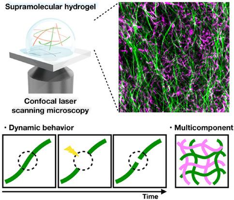
Multicomponent supramolecular hydrogels are promising scaffolds for applications in biosensors and controlled drug release due to their designer stimulus responsiveness. To achieve rational construction of multicomponent supramolecular hydrogel systems, their in‐depth structural analysis is essential but still challenging. Confocal laser scanning microscopy (CLSM) has emerged as a powerful tool for structural analysis of multicomponent supramolecular hydrogels. CLSM imaging enables real‐time observation of the hydrogels without the need of drying and/or freezing to elucidate their static and dynamic properties. Through multiple, selective fluorescent staining of materials of interest, multiple domains formed in supramolecular hydrogels (e. g. inorganic materials and self‐sorting nanofibers) can also be visualized. CLSM and the related microscopic techniques will be indispensable to investigate complex life‐inspired supramolecular chemical systems.
中文翻译:

共聚焦激光扫描显微镜在超分子化学中的功能:刺激反应性多组分超分子水凝胶的原位实时成像。
多组分超分子水凝胶因其设计者的刺激响应性而成为在生物传感器和控制药物释放中应用的有前途的支架。为了实现多组分超分子水凝胶体系的合理构建,它们的深入结构分析是必不可少的,但仍具有挑战性。共聚焦激光扫描显微镜(CLSM)已成为一种用于多组分超分子水凝胶结构分析的强大工具。CLSM成像可实时观察水凝胶,而无需干燥和/或冷冻以阐明其静态和动态特性。通过对目标材料进行多次选择性荧光染色,超分子水凝胶中形成的多个结构域(例如无机材料和自分选纳米纤维)也可以可视化。
更新日期:2020-01-17
中文翻译:
共聚焦激光扫描显微镜在超分子化学中的功能:刺激反应性多组分超分子水凝胶的原位实时成像。
多组分超分子水凝胶因其设计者的刺激响应性而成为在生物传感器和控制药物释放中应用的有前途的支架。为了实现多组分超分子水凝胶体系的合理构建,它们的深入结构分析是必不可少的,但仍具有挑战性。共聚焦激光扫描显微镜(CLSM)已成为一种用于多组分超分子水凝胶结构分析的强大工具。CLSM成像可实时观察水凝胶,而无需干燥和/或冷冻以阐明其静态和动态特性。通过对目标材料进行多次选择性荧光染色,超分子水凝胶中形成的多个结构域(例如无机材料和自分选纳米纤维)也可以可视化。


 京公网安备 11010802027423号