当前位置:
X-MOL 学术
›
Colloids Surf. B Biointerfaces
›
论文详情
Our official English website, www.x-mol.net, welcomes your
feedback! (Note: you will need to create a separate account there.)
Interaction of nitrofurantoin with lipid langmuir monolayers as cellular membrane models distinguished with tensiometry and infrared spectroscopy.
Colloids and Surfaces B: Biointerfaces ( IF 5.4 ) Pub Date : 2020-01-16 , DOI: 10.1016/j.colsurfb.2020.110794 André C Machado 1 , Luciano Caseli 1
Colloids and Surfaces B: Biointerfaces ( IF 5.4 ) Pub Date : 2020-01-16 , DOI: 10.1016/j.colsurfb.2020.110794 André C Machado 1 , Luciano Caseli 1
Affiliation
|
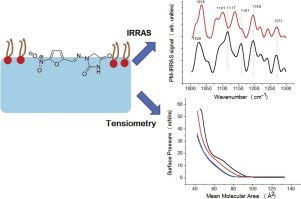
Knowing how a drug interacts with cell membranes is important to understand and predict its effects at the molecular level. Therefore, we aimed to study the interaction of nitrofurantoin (NFT), a compound with potential antibiotic and antitumor properties, with lipidic biological interfaces using Langmuir monolayers. We employed the phospholipids 1,2-dipalmitoyl-sn-glycero-3-phosphocholine (DPPC) and 1,2-dipalmitoyl-sn-glycero-3-phospho-l-serine (DPPS), which were spread on the surface of water to form Langmuir films, to investigate the membrane-drug interactions. The interaction of the drug with the lipid monolayers was evaluated by using surface pressure-area isotherms, surface pressure-time kinetic curves, Brewster angle microscopy (BAM), and polarization-modulated infrared reflection-absorption spectroscopy (PM-IRRAS). Nitrofurantoin shifted the isotherms to lower DPPC molecular areas, indicating monolayer condensation, and to higher DPPS molecular areas, indicating monolayer expansion. Meanwhile, BAM images showed the appearance of interfacial aggregates for DPPS, but not for DPPC, in the presence of NFT. PM-IRRAS spectra showed that bands related to methylene stretches changed their relative intensities and maximum position related to their asymmetric and symmetric modes for both lipids. This suggested an alteration of the monolayer packing degree and the conformational order of the lipid alkyl chains, which were related to an increase in configurational order for DPPS, but disorder for DPPC. In conclusion, NFT caused distinctive changes in the thermodynamic, morphological, and structural properties of DPPC and DPPS monolayers, which may be associated with its bioactivity in cellular membranes and other lipidic interfaces of pharmaceutical interest.
中文翻译:

呋喃妥因与脂质朗缪尔单层的相互作用作为细胞膜模型,具有张力测定法和红外光谱法。
了解药物如何与细胞膜相互作用对于了解和预测其在分子水平的作用非常重要。因此,我们旨在研究使用呋喃单分子膜研究具有潜在抗生素和抗肿瘤特性的化合物呋喃妥因(NFT)与脂质生物界面的相互作用。我们使用了分布在水表面的磷脂1,2-二棕榈酰-sn-甘油-3-磷酸胆碱(DPPC)和1,2-二棕榈酰-sn-甘油--3-磷酸-1-丝氨酸(DPPS)形成Langmuir膜,以研究膜-药物相互作用。通过使用表面压力-面积等温线,表面压力-时间动力学曲线,布鲁斯特角显微镜(BAM)和偏振调制红外反射吸收光谱(PM-IRRAS)评估了药物与脂质单层的相互作用。呋喃妥因将等温线转移到较低的DPPC分子区域,表明单层缩合,而转移到较高的DPPS分子区域,表明单层膨胀。同时,在存在NFT的情况下,BAM图像显示了DPPS的界面聚集体外观,而DPPC则没有。PM-IRRAS光谱表明,与亚甲基拉伸相关的谱带改变了它们的相对强度和最大位置,这与两种脂质的不对称和对称模式有关。这暗示了脂烷基链的单层堆积度和构象顺序的改变,这与DPPS的构象顺序的增加有关,而与DPPC的乱序有关。总之,NFT引起了DPPC和DPPS单层热力学,形态和结构性质的显着变化,
更新日期:2020-01-16
中文翻译:
呋喃妥因与脂质朗缪尔单层的相互作用作为细胞膜模型,具有张力测定法和红外光谱法。
了解药物如何与细胞膜相互作用对于了解和预测其在分子水平的作用非常重要。因此,我们旨在研究使用呋喃单分子膜研究具有潜在抗生素和抗肿瘤特性的化合物呋喃妥因(NFT)与脂质生物界面的相互作用。我们使用了分布在水表面的磷脂1,2-二棕榈酰-sn-甘油-3-磷酸胆碱(DPPC)和1,2-二棕榈酰-sn-甘油--3-磷酸-1-丝氨酸(DPPS)形成Langmuir膜,以研究膜-药物相互作用。通过使用表面压力-面积等温线,表面压力-时间动力学曲线,布鲁斯特角显微镜(BAM)和偏振调制红外反射吸收光谱(PM-IRRAS)评估了药物与脂质单层的相互作用。呋喃妥因将等温线转移到较低的DPPC分子区域,表明单层缩合,而转移到较高的DPPS分子区域,表明单层膨胀。同时,在存在NFT的情况下,BAM图像显示了DPPS的界面聚集体外观,而DPPC则没有。PM-IRRAS光谱表明,与亚甲基拉伸相关的谱带改变了它们的相对强度和最大位置,这与两种脂质的不对称和对称模式有关。这暗示了脂烷基链的单层堆积度和构象顺序的改变,这与DPPS的构象顺序的增加有关,而与DPPC的乱序有关。总之,NFT引起了DPPC和DPPS单层热力学,形态和结构性质的显着变化,


 京公网安备 11010802027423号