Theranostics ( IF 12.4 ) Pub Date : 2020-01-01 , DOI: 10.7150/thno.38501 Wim van Boxtel 1 , Susanne Lütje 2, 3 , Ilse C H van Engen-van Grunsven 4 , Gerald W Verhaegh 5 , Jack A Schalken 5 , Marianne A Jonker 6 , James Nagarajah 2, 7 , Martin Gotthardt 2 , Carla M L van Herpen 1
|
Rationale: Treatment options for recurrent and/or metastatic (R/M) adenoid cystic carcinoma (ACC) and salivary duct carcinoma (SDC), major subtypes of salivary gland cancer, are limited. Both tumors often show overexpression of prostate-specific membrane antigen (PSMA). In prostate cancer, PSMA-ligands labeled with 68Ga or 177Lu are used for imaging and therapy, respectively. Primary aim of this study in R/M ACC and SDC patients was to systematically investigate 68Ga-PSMA-uptake by PET/CT imaging to determine if PSMA radionuclide therapy could be a treatment option.
Methods: In a prospective phase II study, PET/CT imaging was performed 1 h post injection of 68Ga-PSMA-HBED-CC in 15 ACC patients and 10 SDC patients. Maximum standardized uptake values (SUV) were determined in tumor lesions. Immunohistochemical PSMA expression was scored in primary tumors and metastatic tissue. Standard imaging (MRI or CT) was performed for comparison.
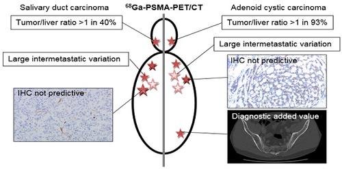
Results: In ACC patients, SUVmax ranged from 1.1 to 30.2 with a tumor/liver-ratio >1 in 13 out of 14 evaluable patients (93%). In SDC patients, SUVmax ranged from 0.3 to 25.9 with a tumor/liver-ratio >1 in 4 out of 10 patients (40%). We found a large intra-patient inter-metastatic variation in uptake of 68Ga-PSMA, and immunohistochemistry did not predict ligand uptake in ACC and SDC. Finally, PSMA-PET detected additional bone metastases compared to CT in 2 ACC patients with unexplained pain.
Conclusion: In 93% of ACC patients and 40% of SDC patients we detected relevant PSMA-ligand uptake, which warrants to study PSMA radionuclide therapy in these patients. Additionally, our data provide arguments for patient selection and treatment timing. Finally, PSMA-PET imaging has added diagnostic value compared to CT in selected patients.
中文翻译:
68Ga-PSMA-HBED-CC PET / CT成像用于腺样囊性癌和涎腺导管癌:2期成像研究。
理由:对于唾液腺癌的主要亚型,复发和/或转移性(R / M)腺样囊性癌(ACC)和唾液管癌(SDC)的治疗选择是有限的。两种肿瘤通常都表现出前列腺特异性膜抗原(PSMA)的过度表达。在前列腺癌中,标记有68 Ga或177 Lu的PSMA配体分别用于成像和治疗。这项研究在R / M ACC和SDC患者中的主要目的是系统地研究PET / CT成像对68 Ga-PSMA的摄取,以确定PSMA放射性核素治疗是否可以作为治疗选择。
方法:在一项前瞻性II期研究中,在15例ACC患者和10例SDC患者中,注射68 Ga-PSMA-HBED-CC后1小时进行了PET / CT成像。在肿瘤病变中确定最大标准化摄取值(SUV)。在原发性肿瘤和转移组织中对免疫组织化学PSMA表达进行评分。进行标准成像(MRI或CT)以进行比较。
结果:在14例可评估患者中,有13例的ACC患者的SUV max在1.1至30.2之间,肿瘤/肝脏比率> 1。在SDC患者中,SUV最大范围为0.3至25.9,其中肿瘤/肝比率> 1的患者中有十分之四(40%)。我们发现患者体内68 Ga-PSMA的摄取之间存在较大的转移间差异,并且免疫组织化学未预测ACC和SDC中的配体摄取。最后,与2例ACC伴有无法解释的疼痛的CT相比,PSMA-PET检测到了更多的骨转移。
结论:在93%的ACC患者和40%的SDC患者中,我们检测到相关的PSMA配体摄取,因此有必要对这些患者进行PSMA放射性核素治疗研究。此外,我们的数据为患者选择和治疗时机提供了依据。最后,与CT相比,PSMA-PET成像对某些患者具有更高的诊断价值。


 京公网安备 11010802027423号