当前位置:
X-MOL 学术
›
ACS Biomater. Sci. Eng.
›
论文详情
Our official English website, www.x-mol.net, welcomes your
feedback! (Note: you will need to create a separate account there.)
3D Printing of Vascular Tubes Using Bioelastomer Prepolymers by Freeform Reversible Embedding
ACS Biomaterials Science & Engineering ( IF 5.4 ) Pub Date : 2020-02-11 , DOI: 10.1021/acsbiomaterials.9b00676 Houman Savoji 1, 2 , Locke Davenport Huyer 1, 2, 3 , Mohammad Hossein Mohammadi 2, 3 , Benjamin Fook Lun Lai 1 , Naimeh Rafatian 2 , Dawn Bannerman 1, 2, 3 , Mohammad Shoaib 3 , Erin R. Bobicki 3 , Arun Ramachandran 3 , Milica Radisic 1, 2, 3
ACS Biomaterials Science & Engineering ( IF 5.4 ) Pub Date : 2020-02-11 , DOI: 10.1021/acsbiomaterials.9b00676 Houman Savoji 1, 2 , Locke Davenport Huyer 1, 2, 3 , Mohammad Hossein Mohammadi 2, 3 , Benjamin Fook Lun Lai 1 , Naimeh Rafatian 2 , Dawn Bannerman 1, 2, 3 , Mohammad Shoaib 3 , Erin R. Bobicki 3 , Arun Ramachandran 3 , Milica Radisic 1, 2, 3
Affiliation
|
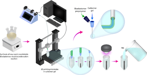
Bioelastomers have been extensively used in tissue engineering applications because of favorable mechanical stability, tunable properties, and chemical versatility. As these materials generally possess low elastic modulus and relatively long gelation time, it is challenging to 3D print them using traditional techniques. Instead, the field of 3D printing has focused preferentially on hydrogels and rigid polyester materials. To develop a versatile approach for 3D printing of elastomers, we used freeform reversible embedding of suspended prepolymers. A family of novel fast photocrosslinakble bioelastomer prepolymers were synthesized from dimethyl itaconate, 1,8-octanediol, and triethyl citrate. Tensile testing confirmed their elastic properties with Young’s moduli in the range of 11–53 kPa. These materials supported cultivation of viable cells and enabled adhesion and proliferation of human umbilical vein endothelial cells. Tubular structures were created by embedding the 3D printed microtubes within a secondary hydrogel that served as a temporary support. Upon photocrosslinking and porogen leaching, the polymers were permeable to small molecules (TRITC-dextran). The polymer microtubes were assembled on the 96-well plates custom made by hot-embossing, as a tool to connect multiple organs-on-a-chip. The endothelialization of the tubes was performed to confirm that these microtubes can be utilized as vascular tubes to support parenchymal tissues seeded on them.
中文翻译:

使用生物弹性体预聚物通过自由形式可逆嵌入进行3D打印血管
由于具有良好的机械稳定性,可调性和化学通用性,生物弹性体已广泛用于组织工程应用。由于这些材料通常具有较低的弹性模量和相对较长的胶凝时间,因此使用传统技术对其进行3D打印具有挑战性。相反,3D打印领域优先关注水凝胶和硬质聚酯材料。为了开发一种用于弹性体3D打印的通用方法,我们使用了悬浮预聚物的自由形式可逆嵌入。从衣康酸二甲酯,1,8-辛二醇和柠檬酸三乙酯合成了一系列新型的快速可光交联的生物弹性体预聚物。拉伸测试证实了它们的弹性,其杨氏模量在11–53 kPa范围内。这些材料支持活细胞的培养,并使人脐静脉内皮细胞粘附和增殖。通过将3D打印的微管嵌入到用作临时支撑物的辅助水凝胶中来创建管状结构。光交联和致孔剂浸出后,聚合物可渗透小分子(TRITC-葡聚糖)。将聚合物微管组装在通过热压印定制的96孔板上,作为连接多个芯片上芯片的工具。进行管的内皮化以确认这些微管可用作血管管以支持接种在其上的实质组织。通过将3D打印的微管嵌入到用作临时支撑物的辅助水凝胶中来创建管状结构。光交联和致孔剂浸出后,聚合物可渗透小分子(TRITC-葡聚糖)。将聚合物微管组装在通过热压印定制的96孔板上,作为连接多个芯片上芯片的工具。进行管的内皮化以确认这些微管可用作血管管以支持接种在其上的实质组织。通过将3D打印的微管嵌入到用作临时支撑物的辅助水凝胶中来创建管状结构。光交联和致孔剂浸出后,聚合物可渗透小分子(TRITC-葡聚糖)。将聚合物微管组装在通过热压印定制的96孔板上,作为连接多个芯片上芯片的工具。进行管的内皮化以确认这些微管可用作血管管以支持接种在其上的实质组织。
更新日期:2020-02-11
中文翻译:
使用生物弹性体预聚物通过自由形式可逆嵌入进行3D打印血管
由于具有良好的机械稳定性,可调性和化学通用性,生物弹性体已广泛用于组织工程应用。由于这些材料通常具有较低的弹性模量和相对较长的胶凝时间,因此使用传统技术对其进行3D打印具有挑战性。相反,3D打印领域优先关注水凝胶和硬质聚酯材料。为了开发一种用于弹性体3D打印的通用方法,我们使用了悬浮预聚物的自由形式可逆嵌入。从衣康酸二甲酯,1,8-辛二醇和柠檬酸三乙酯合成了一系列新型的快速可光交联的生物弹性体预聚物。拉伸测试证实了它们的弹性,其杨氏模量在11–53 kPa范围内。这些材料支持活细胞的培养,并使人脐静脉内皮细胞粘附和增殖。通过将3D打印的微管嵌入到用作临时支撑物的辅助水凝胶中来创建管状结构。光交联和致孔剂浸出后,聚合物可渗透小分子(TRITC-葡聚糖)。将聚合物微管组装在通过热压印定制的96孔板上,作为连接多个芯片上芯片的工具。进行管的内皮化以确认这些微管可用作血管管以支持接种在其上的实质组织。通过将3D打印的微管嵌入到用作临时支撑物的辅助水凝胶中来创建管状结构。光交联和致孔剂浸出后,聚合物可渗透小分子(TRITC-葡聚糖)。将聚合物微管组装在通过热压印定制的96孔板上,作为连接多个芯片上芯片的工具。进行管的内皮化以确认这些微管可用作血管管以支持接种在其上的实质组织。通过将3D打印的微管嵌入到用作临时支撑物的辅助水凝胶中来创建管状结构。光交联和致孔剂浸出后,聚合物可渗透小分子(TRITC-葡聚糖)。将聚合物微管组装在通过热压印定制的96孔板上,作为连接多个芯片上芯片的工具。进行管的内皮化以确认这些微管可用作血管管以支持接种在其上的实质组织。


 京公网安备 11010802027423号