当前位置:
X-MOL 学术
›
Cell Death Dis.
›
论文详情
Our official English website, www.x-mol.net, welcomes your
feedback! (Note: you will need to create a separate account there.)
Drp1-mediated mitochondrial fission promotes renal fibroblast activation and fibrogenesis.
Cell Death & Disease ( IF 8.1 ) Pub Date : 2020-01-16 , DOI: 10.1038/s41419-019-2218-5 Yating Wang 1, 2, 3 , Miaoqing Lu 2, 3, 4 , Liping Xiong 1, 2, 3 , Jinjin Fan 1, 2, 3 , Yi Zhou 1, 2, 3 , Huiyan Li 1, 2, 3 , Xuan Peng 1, 2, 3 , Zhong Zhong 1, 2, 3 , Yihan Wang 5 , Fengxian Huang 1, 2, 3 , Wei Chen 1, 2, 3 , Xueqing Yu 1, 2, 3 , Haiping Mao 1, 2, 3
Cell Death & Disease ( IF 8.1 ) Pub Date : 2020-01-16 , DOI: 10.1038/s41419-019-2218-5 Yating Wang 1, 2, 3 , Miaoqing Lu 2, 3, 4 , Liping Xiong 1, 2, 3 , Jinjin Fan 1, 2, 3 , Yi Zhou 1, 2, 3 , Huiyan Li 1, 2, 3 , Xuan Peng 1, 2, 3 , Zhong Zhong 1, 2, 3 , Yihan Wang 5 , Fengxian Huang 1, 2, 3 , Wei Chen 1, 2, 3 , Xueqing Yu 1, 2, 3 , Haiping Mao 1, 2, 3
Affiliation
|
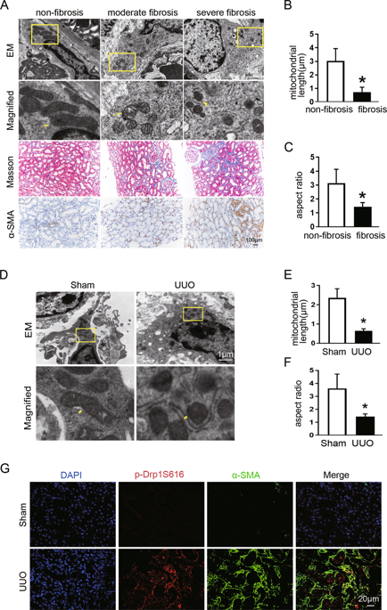
Excessive mitochondrial fission acts as a pro-proliferative marker in some cancers and organ fibrosis; its potential role in renal fibroblast activation and fibrogenesis has never been investigated. Here, we showed more pronounced fragmented mitochondria in fibrotic than in non-fibrotic renal fibroblast in humans and mice. In a mouse model of obstructive nephropathy, phosphorylation of Drp1 at serine 616 (p-Drp1S616) and acetylation of H3K27(H3K27ac) was increased in fibrotic kidneys; pharmacological inhibition of mitochondrial fission by mdivi-1 substantially reduced H3K27ac levels, fibroblasts accumulation, and interstitial fibrosis. Moreover, mdivi-1 treatment was able to attenuate the established renal fibrosis. In cultured renal interstitial fibroblasts, targeting Drp1 using pharmacological inhibitor or siRNA suppressed TGF-β1-elicited cell activation and proliferation, as evidenced by inhibiting expression of α-smooth muscle actin (α-SMA) and collagen I, as well as by reducing DNA synthesis. In contrast, Drp1 deletion enhanced cell apoptosis, along with decreased mitochondrial fragmentation, mtROS elevation, and glycolytic shift upon TGF-β1 stimulation. In Drp1 deletion fibroblasts, re-expression of wild-type Drp1 rather than Drp1S616A mutant restores the reduction of TGF-β-induced-Drp1 phosphorylation, H3K27ac, and cell activation. Moreover, TGF-β1 treatment increased the enrichment of H3K27ac at the promoters of α-SMA and PCNA, which was reversed in Drp1-knockdown fibroblasts co-transfected with empty vector or Drp1S616A, but not wild-type Drp1. Collectively, our results imply that inhibiting p-Drp1S616-mediated mitochondrial fission attenuates fibroblast activation and proliferation in renal fibrosis through epigenetic regulation of fibrosis-related genes transcription and may serve as a therapeutic target for retarding progression of chronic kidney disease.
中文翻译:

Drp1介导的线粒体裂变促进肾脏成纤维细胞活化和纤维化。
线粒体过度分裂是某些癌症和器官纤维化中增殖的标志。尚未研究其在肾成纤维细胞活化和纤维形成中的潜在作用。在这里,我们在人类和小鼠的纤维化中显示出比非纤维化性肾成纤维细胞中更明显的线粒体碎片。在阻塞性肾病的小鼠模型中,在纤维化肾脏中,丝氨酸616处Drp1的磷酸化(p-Drp1S616)和H3K27(H3K27ac)的乙酰化增加;mdivi-1对线粒体裂变的药理学抑制作用显着降低了H3K27ac水平,成纤维细胞积累和间质纤维化。此外,mdivi-1治疗能够减轻已建立的肾纤维化。在培养的肾间质成纤维细胞中 使用药理抑制剂或siRNA靶向Drp1可抑制TGF-β1诱导的细胞活化和增殖,这可通过抑制α平滑肌肌动蛋白(α-SMA)和胶原I的表达以及减少DNA合成来证明。相反,Drp1缺失增强了细胞凋亡,并降低了线粒体碎片,mtROS升高和TGF-β1刺激后的糖酵解位移。在Drp1缺失成纤维细胞中,野生型Drp1而不是Drp1S616A突变体的重新表达恢复了TGF-β诱导的Drp1磷酸化,H3K27ac和细胞活化的减少。此外,TGF-β1处理增加了H3K27ac在α-SMA和PCNA启动子上的富集,这在与空载体或Drp1S616A共转染的Drp1-敲低成纤维细胞中是相反的,但在野生型Drp1中却没有。总的来说,
更新日期:2020-01-16
中文翻译:
Drp1介导的线粒体裂变促进肾脏成纤维细胞活化和纤维化。
线粒体过度分裂是某些癌症和器官纤维化中增殖的标志。尚未研究其在肾成纤维细胞活化和纤维形成中的潜在作用。在这里,我们在人类和小鼠的纤维化中显示出比非纤维化性肾成纤维细胞中更明显的线粒体碎片。在阻塞性肾病的小鼠模型中,在纤维化肾脏中,丝氨酸616处Drp1的磷酸化(p-Drp1S616)和H3K27(H3K27ac)的乙酰化增加;mdivi-1对线粒体裂变的药理学抑制作用显着降低了H3K27ac水平,成纤维细胞积累和间质纤维化。此外,mdivi-1治疗能够减轻已建立的肾纤维化。在培养的肾间质成纤维细胞中 使用药理抑制剂或siRNA靶向Drp1可抑制TGF-β1诱导的细胞活化和增殖,这可通过抑制α平滑肌肌动蛋白(α-SMA)和胶原I的表达以及减少DNA合成来证明。相反,Drp1缺失增强了细胞凋亡,并降低了线粒体碎片,mtROS升高和TGF-β1刺激后的糖酵解位移。在Drp1缺失成纤维细胞中,野生型Drp1而不是Drp1S616A突变体的重新表达恢复了TGF-β诱导的Drp1磷酸化,H3K27ac和细胞活化的减少。此外,TGF-β1处理增加了H3K27ac在α-SMA和PCNA启动子上的富集,这在与空载体或Drp1S616A共转染的Drp1-敲低成纤维细胞中是相反的,但在野生型Drp1中却没有。总的来说,


 京公网安备 11010802027423号