JAMA Dermatology ( IF 11.5 ) Pub Date : 2020-01-15 , DOI: 10.1001/jamadermatol.2019.4274 Alessandro Di Stefani 1, 2 , Simone Cappilli 1, 2 , Ketty Peris 1, 2
|
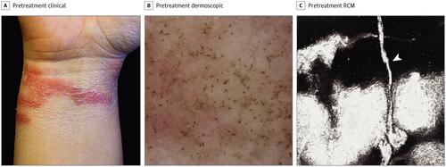
This case report describes the utility of dermoscopy and reflectance confocal microscopy (RCM) in confirming clinical diagnosis and monitoring treatment response in patients with jellyfish stings. Jellyfish stings are a common nuisance for sea bathers worldwide. Pelagia noctiluca is usually considered to be the most important species of jellyfish in the Mediterranean Sea owing to its widespread distribution, ecological role, and accidental interactions with humans.1 The common mechanism through which jellyfish discharge toxins during contact with their prey is attributed to structures called nematocysts, which are barbs studding the tentacles as well as the upper surface of the bell.1
中文翻译:
用于诊断和治疗监测的水母St体内成像。
该病例报告描述了皮肤镜检查和反射共聚焦显微镜(RCM)在确认临床诊断和监测水母st伤患者治疗反应中的效用。水母ing是全世界海水浴者的常见困扰。由于其广泛分布,生态作用以及与人类的偶然相互作用,因此夜蛾通常被认为是地中海中最重要的水母种类。1水母在与猎物接触时释放毒素的常见机制归因于称为线虫囊的结构,该结构是倒钩触角和钟形上表面的倒钩。1个


 京公网安备 11010802027423号