当前位置:
X-MOL 学术
›
Anal. Chem.
›
论文详情
Our official English website, www.x-mol.net, welcomes your
feedback! (Note: you will need to create a separate account there.)
Injection Molded Microfluidics for Establishing High-Density Single Cell Arrays in an Open Hydrogel Format.
Analytical Chemistry ( IF 6.7 ) Pub Date : 2020-01-14 , DOI: 10.1021/acs.analchem.9b05099 Ying Li 1, 2 , Jeffrey D Motschman 1 , Sean T Kelly 1 , Benjamin B Yellen 1, 3
Analytical Chemistry ( IF 6.7 ) Pub Date : 2020-01-14 , DOI: 10.1021/acs.analchem.9b05099 Ying Li 1, 2 , Jeffrey D Motschman 1 , Sean T Kelly 1 , Benjamin B Yellen 1, 3
Affiliation
|
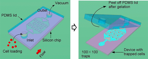
Here, we develop an injection molded microfluidic approach for single cell analysis by making use of (1) rapidly curing injectable hydrogels, (2) a high density microfluidic weir trap array, and (3) reversibly bonded PDMS lids that are strong enough to withstand the injection molding process, but which can be peeled off after the hydrogel sets. This approach allows for single cell patterns to be created with densities exceeding 40 cells per mm2, is amenable to high speed imaging, and creates microfluidic devices that enable efficient nutrient and gas exchange and the delivery of specific biological and chemical reagents to individual cells. We show that it is possible to organize up to 10 000 single cells in a few minutes on the device, and we developed an image analysis program to automatically analyze the single-cell capture efficiency. The results show single cell trapping rates were better than 80%. We also demonstrate that the genomic DNA of the single cells trapped in the hydrogel can be amplified via localized, multiple displacement amplification in a massively parallel format, which offers a promising strategy for analyzing single cell genomes. Finally, we show the ability to perform selective staining of individual cells with a commercial bioprinter, providing proof of concept of its ability to deliver tailored reagents to specific cells in an array for future downstream analysis. This injection molded microfluidic approach leverages the benefits of both closed and open microfluidics, allows multiday single cell cultures, direct access to the trapped cells for genotypic end point studies.
中文翻译:

用于以开放水凝胶形式建立高密度单细胞阵列的注塑微流体。
在这里,我们开发了一种用于单细胞分析的注射成型微流体方法,利用(1)快速固化的可注射水凝胶,(2)高密度微流体堰陷阱阵列,以及(3)强度足以承受的可逆结合的PDMS盖注射成型工艺,但在水凝胶凝固后可以剥离。这种方法允许创建密度超过每平方毫米 40 个细胞的单细胞图案,适合高速成像,并创建微流体装置,能够实现有效的营养物和气体交换以及将特定的生物和化学试剂输送到单个细胞。我们证明可以在几分钟内在设备上组织多达 10 000 个单细胞,并且我们开发了一种图像分析程序来自动分析单细胞捕获效率。结果显示单细胞捕获率优于80%。我们还证明,水凝胶中捕获的单细胞的基因组 DNA 可以通过大规模并行格式的局部多重置换扩增进行扩增,这为分析单细胞基因组提供了一种有前途的策略。最后,我们展示了使用商业生物打印机对单个细胞进行选择性染色的能力,提供了其向阵列中的特定细胞提供定制试剂以供未来下游分析的能力的概念证明。这种注塑微流体方法利用了封闭式和开放式微流体的优点,允许进行多天的单细胞培养,直接接触捕获的细胞进行基因型终点研究。
更新日期:2020-01-14
中文翻译:
用于以开放水凝胶形式建立高密度单细胞阵列的注塑微流体。
在这里,我们开发了一种用于单细胞分析的注射成型微流体方法,利用(1)快速固化的可注射水凝胶,(2)高密度微流体堰陷阱阵列,以及(3)强度足以承受的可逆结合的PDMS盖注射成型工艺,但在水凝胶凝固后可以剥离。这种方法允许创建密度超过每平方毫米 40 个细胞的单细胞图案,适合高速成像,并创建微流体装置,能够实现有效的营养物和气体交换以及将特定的生物和化学试剂输送到单个细胞。我们证明可以在几分钟内在设备上组织多达 10 000 个单细胞,并且我们开发了一种图像分析程序来自动分析单细胞捕获效率。结果显示单细胞捕获率优于80%。我们还证明,水凝胶中捕获的单细胞的基因组 DNA 可以通过大规模并行格式的局部多重置换扩增进行扩增,这为分析单细胞基因组提供了一种有前途的策略。最后,我们展示了使用商业生物打印机对单个细胞进行选择性染色的能力,提供了其向阵列中的特定细胞提供定制试剂以供未来下游分析的能力的概念证明。这种注塑微流体方法利用了封闭式和开放式微流体的优点,允许进行多天的单细胞培养,直接接触捕获的细胞进行基因型终点研究。


 京公网安备 11010802027423号