Our official English website, www.x-mol.net, welcomes your feedback! (Note: you will need to create a separate account there.)
Super-Resolution Mapping of Single Nanoparticles inside Tumor Spheroids.
Small ( IF 13.3 ) Pub Date : 2020-01-14 , DOI: 10.1002/smll.201905572 Yongtao Liu 1 , Fan Wang 1 , Hongxu Lu 2 , Guocheng Fang 1 , Shihui Wen 1 , Chaohao Chen 1 , Xuchen Shan 1 , Xiaoxue Xu 1 , Lin Zhang 2 , Martina Stenzel 2 , Dayong Jin 1, 3
Small ( IF 13.3 ) Pub Date : 2020-01-14 , DOI: 10.1002/smll.201905572 Yongtao Liu 1 , Fan Wang 1 , Hongxu Lu 2 , Guocheng Fang 1 , Shihui Wen 1 , Chaohao Chen 1 , Xuchen Shan 1 , Xiaoxue Xu 1 , Lin Zhang 2 , Martina Stenzel 2 , Dayong Jin 1, 3
Affiliation
|
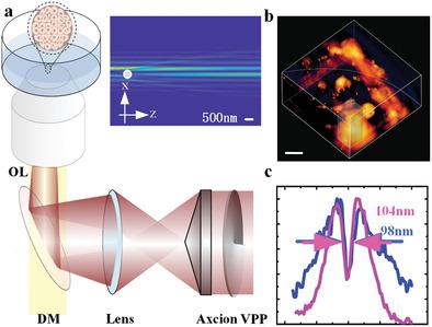
Cancer spheroids have structural, functional, and physiological similarities to the tumor, and have become a low-cost in vitro model to study the physiological responses of single cells and therapeutic efficacy of drugs. However, the tiny spheroid, made of a cluster of high-density cells, is highly scattering and absorptive, which prevents light microscopy techniques to reach the depth inside spheroids with high resolution. Here, a method is reported for super-resolution mapping of single nanoparticles inside a spheroid. It first takes advantage of the self-healing property of a "nondiffractive" doughnut-shaped Bessel beam from a 980 nm diode laser as the excitation, and further employs the nonlinear response of the 800 nm emission from upconversion nanoparticles, so that both excitation and emission at the near-infrared can experience minimal loss through the spheroid. These strategies lead to the development of a new nanoscopy modality with a resolution of 37 nm, 1/26th of the excitation wavelength. This method enables mapping of single nanoparticles located 55 µm inside a spheroid, with a resolution of 98 nm. It suggests a solution to track single nanoparticles and monitor their release of drugs in 3D multicellar environments.
中文翻译:

肿瘤球体内单个纳米粒子的超分辨率映射。
癌症球体与肿瘤具有结构,功能和生理上的相似性,并已成为研究单个细胞的生理反应和药物治疗功效的低成本体外模型。但是,由一群高密度细胞组成的微小球状体具有很高的散射性和吸收性,这阻止了光学显微镜技术以高分辨率到达球状体内部的深度。在此,报道了一种用于球体内部单个纳米粒子超分辨率映射的方法。它首先利用来自980 nm二极管激光器的“非衍射”甜甜圈形贝塞尔光束的自愈特性作为激发,并进一步利用了上转换纳米粒子发出的800 nm发射的非线性响应,因此,近红外光的激发和发射都可以通过球体损失最小。这些策略导致了新的纳米显微镜形式的发展,其分辨率为37 nm,是激发波长的1/26。这种方法可以绘制位于球体内55 µm的单个纳米颗粒,分辨率为98 nm。它提出了一种跟踪单个纳米颗粒并监控3D多腔环境中药物释放的解决方案。
更新日期:2020-02-13
中文翻译:
肿瘤球体内单个纳米粒子的超分辨率映射。
癌症球体与肿瘤具有结构,功能和生理上的相似性,并已成为研究单个细胞的生理反应和药物治疗功效的低成本体外模型。但是,由一群高密度细胞组成的微小球状体具有很高的散射性和吸收性,这阻止了光学显微镜技术以高分辨率到达球状体内部的深度。在此,报道了一种用于球体内部单个纳米粒子超分辨率映射的方法。它首先利用来自980 nm二极管激光器的“非衍射”甜甜圈形贝塞尔光束的自愈特性作为激发,并进一步利用了上转换纳米粒子发出的800 nm发射的非线性响应,因此,近红外光的激发和发射都可以通过球体损失最小。这些策略导致了新的纳米显微镜形式的发展,其分辨率为37 nm,是激发波长的1/26。这种方法可以绘制位于球体内55 µm的单个纳米颗粒,分辨率为98 nm。它提出了一种跟踪单个纳米颗粒并监控3D多腔环境中药物释放的解决方案。


 京公网安备 11010802027423号