当前位置:
X-MOL 学术
›
Int. J. Oral Sci.
›
论文详情
Our official English website, www.x-mol.net, welcomes your
feedback! (Note: you will need to create a separate account there.)
The dimension and morphology of alveolar bone at maxillary anterior teeth in periodontitis: a retrospective analysis-using CBCT.
International Journal of Oral Science ( IF 10.8 ) Pub Date : 2020-01-14 , DOI: 10.1038/s41368-019-0071-0 Xue Zhang 1 , Yuchao Li 1 , Ziming Ge 1 , Haijiao Zhao 1 , Lei Miao 1 , Yaping Pan 1
International Journal of Oral Science ( IF 10.8 ) Pub Date : 2020-01-14 , DOI: 10.1038/s41368-019-0071-0 Xue Zhang 1 , Yuchao Li 1 , Ziming Ge 1 , Haijiao Zhao 1 , Lei Miao 1 , Yaping Pan 1
Affiliation
|
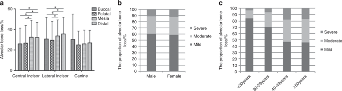
The morphology of the alveolar bone at the maxillary anterior teeth in periodontitis patients was evaluated by cone-beam computed tomography (CBCT) to investigate the distribution of alveolar defects and provide guidance for clinical practice. Ninety periodontitis patients and 30 periodontally healthy individuals were selected to determine the morphology of the alveolar bone at the maxillary anterior teeth according to the degree of bone loss, tooth type, sex and age. The differences in the dimensions between periodontitis patients and healthy individuals were compared, and the distribution of alveolar bone defects was analyzed. A classification system was established regarding the sagittal positions and angulations of the teeth. The buccal residual bone was thicker and the lingual bone was thinner in the periodontitis patients than in the periodontally healthy individuals, and there were differences between the different tooth types, sexes and age subgroups. The buccal undercut was close to the alveolar ridge, while fenestration was reduced and the apical bone height was higher in periodontitis patients than in periodontally healthy individuals. The apical bone height increased with the aggravation of bone loss and age. The proportions of different sagittal positions changed with the aggravation of bone loss. Moreover, the teeth moved more buccally regarding the positions of the maxillary anterior teeth. The morphology of the alveolar bone at the maxillary anterior teeth differed between periodontitis patients and healthy individuals, and the differences were related to the degree of bone loss, tooth type, sex and age.
中文翻译:

牙周炎上颌前牙牙槽骨的尺寸和形态:使用CBCT的回顾性分析。
通过锥束计算机断层扫描(CBCT)评估牙周炎患者上颌前牙的牙槽骨形态,以调查牙槽缺损的分布并为临床实践提供指导。根据骨质流失的程度,类型,性别和年龄,选择了90名牙周炎患者和30名牙周健康患者,以确定上颌前牙的牙槽骨形态。比较牙周炎患者和健康个体在尺寸上的差异,并分析牙槽骨缺损的分布。建立了关于矢状位和牙齿角度的分类系统。与牙周健康个体相比,牙周炎患者的颊侧残留骨更厚,舌骨更薄,并且不同牙齿类型,性别和年龄组之间存在差异。与牙周健康者相比,牙周炎患者的颊侧咬边靠近牙槽,而开窗率降低且根尖骨高度更高。随着骨质流失和年龄的增加,根尖骨高度增加。矢状位的比例随着骨质流失的加剧而变化。此外,就上颌前牙的位置而言,牙齿的颊侧移动更多。牙周炎患者和健康个体的上颌前牙的牙槽骨形态不同,
更新日期:2020-01-14
中文翻译:
牙周炎上颌前牙牙槽骨的尺寸和形态:使用CBCT的回顾性分析。
通过锥束计算机断层扫描(CBCT)评估牙周炎患者上颌前牙的牙槽骨形态,以调查牙槽缺损的分布并为临床实践提供指导。根据骨质流失的程度,类型,性别和年龄,选择了90名牙周炎患者和30名牙周健康患者,以确定上颌前牙的牙槽骨形态。比较牙周炎患者和健康个体在尺寸上的差异,并分析牙槽骨缺损的分布。建立了关于矢状位和牙齿角度的分类系统。与牙周健康个体相比,牙周炎患者的颊侧残留骨更厚,舌骨更薄,并且不同牙齿类型,性别和年龄组之间存在差异。与牙周健康者相比,牙周炎患者的颊侧咬边靠近牙槽,而开窗率降低且根尖骨高度更高。随着骨质流失和年龄的增加,根尖骨高度增加。矢状位的比例随着骨质流失的加剧而变化。此外,就上颌前牙的位置而言,牙齿的颊侧移动更多。牙周炎患者和健康个体的上颌前牙的牙槽骨形态不同,


 京公网安备 11010802027423号