当前位置:
X-MOL 学术
›
NeuroImage
›
论文详情
Our official English website, www.x-mol.net, welcomes your
feedback! (Note: you will need to create a separate account there.)
The dot-compartment revealed? Diffusion MRI with ultra-strong gradients and spherical tensor encoding in the living human brain
NeuroImage ( IF 4.7 ) Pub Date : 2020-04-01 , DOI: 10.1016/j.neuroimage.2020.116534 Chantal M W Tax 1 , Filip Szczepankiewicz 2 , Markus Nilsson 3 , Derek K Jones 4
NeuroImage ( IF 4.7 ) Pub Date : 2020-04-01 , DOI: 10.1016/j.neuroimage.2020.116534 Chantal M W Tax 1 , Filip Szczepankiewicz 2 , Markus Nilsson 3 , Derek K Jones 4
Affiliation
|
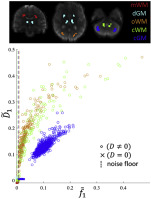
The so-called “dot-compartment” is conjectured in diffusion MRI to represent small spherical spaces, such as cell bodies, in which the diffusion is restricted in all directions. Previous investigations inferred its existence from data acquired with directional diffusion encoding which does not permit a straightforward separation of signals from ‘sticks’ (axons) and signals from ‘dots’. Here we combine isotropic diffusion encoding with ultra-strong diffusion gradients (240 mT/m) to achieve high diffusion-weightings with high signal to noise ratio, while suppressing signal arising from anisotropic water compartments with significant mobility along at least one axis (e.g., axons). A dot-compartment, defined to have apparent diffusion coefficient equal to zero and no exchange, would result in a non-decaying signal at very high b-values (b≳7000s/mm2). With this unique experimental setup, a residual yet slowly decaying signal above the noise floor for b-values as high as 15000s/mm2 was seen clearly in the cerebellar grey matter (GM), and in several white matter (WM) regions to some extent. Upper limits of the dot-signal-fraction were estimated to be 1.8% in cerebellar GM and 0.5% in WM. By relaxing the assumption of zero diffusivity, the signal at high b-values in cerebellar GM could be represented more accurately by an isotropic water pool with a low apparent diffusivity of 0.12 μm2/ms and a substantial signal fraction of 9.7%. The T2 of this component was estimated to be around 61ms. This remaining signal at high b-values has potential to serve as a novel and simple marker for isotropically-restricted water compartments in cerebellar GM.
中文翻译:

点隔间显露出来?在活人脑中具有超强梯度和球形张量编码的扩散 MRI
所谓的“点室”在扩散MRI中被推测为代表小的球形空间,例如细胞体,其中向各个方向的扩散都受到限制。先前的研究从使用定向扩散编码获得的数据推断其存在,这不允许直接分离来自“棒”(轴突)的信号和来自“点”的信号。在这里,我们将各向同性扩散编码与超强扩散梯度 (240 mT/m) 相结合,以实现具有高信噪比的高扩散权重,同时抑制沿至少一个轴具有显着流动性的各向异性水隔室产生的信号(例如,轴突)。定义为表观扩散系数为零且无交换的点隔室将在非常高的 b 值 (b≳7000s/mm2) 下产生非衰减信号。通过这种独特的实验设置,在小脑灰质 (GM) 和一些白质 (WM) 区域中可以清楚地看到 b 值高达 15000s/mm2 的本底噪声之上的残留但缓慢衰减的信号. 点信号分数的上限估计在小脑 GM 中为 1.8%,在 WM 中为 0.5%。通过放宽零扩散率的假设,小脑 GM 中高 b 值的信号可以更准确地由各向同性水池表示,该水池具有 0.12 μm2/ms 的低表观扩散率和 9.7% 的大量信号分数。该组件的 T2 估计约为 61 毫秒。这种在高 b 值下的剩余信号有可能作为小脑 GM 中各向同性限制水区室的新颖而简单的标记。在小脑灰质 (GM) 和一些白质 (WM) 区域中,可以清楚地看到 b 值高达 15000s/mm2 的本底噪声之上的残留但缓慢衰减的信号。点信号分数的上限估计在小脑 GM 中为 1.8%,在 WM 中为 0.5%。通过放宽零扩散率的假设,小脑 GM 中高 b 值的信号可以更准确地由各向同性水池表示,该水池具有 0.12 μm2/ms 的低表观扩散率和 9.7% 的大量信号分数。该组件的 T2 估计约为 61 毫秒。这种在高 b 值下的剩余信号有可能作为小脑 GM 中各向同性限制水区室的新颖而简单的标记。在小脑灰质 (GM) 和一些白质 (WM) 区域中,可以清楚地看到 b 值高达 15000s/mm2 的本底噪声之上的残留但缓慢衰减的信号。点信号分数的上限估计在小脑 GM 中为 1.8%,在 WM 中为 0.5%。通过放宽零扩散率的假设,小脑 GM 中高 b 值的信号可以更准确地由各向同性水池表示,该水池具有 0.12 μm2/ms 的低表观扩散率和 9.7% 的大量信号分数。该组件的 T2 估计约为 61 毫秒。这种在高 b 值下的剩余信号有可能作为小脑 GM 中各向同性限制水区室的新颖而简单的标记。
更新日期:2020-04-01
中文翻译:
点隔间显露出来?在活人脑中具有超强梯度和球形张量编码的扩散 MRI
所谓的“点室”在扩散MRI中被推测为代表小的球形空间,例如细胞体,其中向各个方向的扩散都受到限制。先前的研究从使用定向扩散编码获得的数据推断其存在,这不允许直接分离来自“棒”(轴突)的信号和来自“点”的信号。在这里,我们将各向同性扩散编码与超强扩散梯度 (240 mT/m) 相结合,以实现具有高信噪比的高扩散权重,同时抑制沿至少一个轴具有显着流动性的各向异性水隔室产生的信号(例如,轴突)。定义为表观扩散系数为零且无交换的点隔室将在非常高的 b 值 (b≳7000s/mm2) 下产生非衰减信号。通过这种独特的实验设置,在小脑灰质 (GM) 和一些白质 (WM) 区域中可以清楚地看到 b 值高达 15000s/mm2 的本底噪声之上的残留但缓慢衰减的信号. 点信号分数的上限估计在小脑 GM 中为 1.8%,在 WM 中为 0.5%。通过放宽零扩散率的假设,小脑 GM 中高 b 值的信号可以更准确地由各向同性水池表示,该水池具有 0.12 μm2/ms 的低表观扩散率和 9.7% 的大量信号分数。该组件的 T2 估计约为 61 毫秒。这种在高 b 值下的剩余信号有可能作为小脑 GM 中各向同性限制水区室的新颖而简单的标记。在小脑灰质 (GM) 和一些白质 (WM) 区域中,可以清楚地看到 b 值高达 15000s/mm2 的本底噪声之上的残留但缓慢衰减的信号。点信号分数的上限估计在小脑 GM 中为 1.8%,在 WM 中为 0.5%。通过放宽零扩散率的假设,小脑 GM 中高 b 值的信号可以更准确地由各向同性水池表示,该水池具有 0.12 μm2/ms 的低表观扩散率和 9.7% 的大量信号分数。该组件的 T2 估计约为 61 毫秒。这种在高 b 值下的剩余信号有可能作为小脑 GM 中各向同性限制水区室的新颖而简单的标记。在小脑灰质 (GM) 和一些白质 (WM) 区域中,可以清楚地看到 b 值高达 15000s/mm2 的本底噪声之上的残留但缓慢衰减的信号。点信号分数的上限估计在小脑 GM 中为 1.8%,在 WM 中为 0.5%。通过放宽零扩散率的假设,小脑 GM 中高 b 值的信号可以更准确地由各向同性水池表示,该水池具有 0.12 μm2/ms 的低表观扩散率和 9.7% 的大量信号分数。该组件的 T2 估计约为 61 毫秒。这种在高 b 值下的剩余信号有可能作为小脑 GM 中各向同性限制水区室的新颖而简单的标记。


 京公网安备 11010802027423号