当前位置:
X-MOL 学术
›
ACS Chem. Neurosci.
›
论文详情
Our official English website, www.x-mol.net, welcomes your
feedback! (Note: you will need to create a separate account there.)
Differentiation among Glioblastomas, Primary Cerebral Lymphomas, and Solitary Brain Metastases Using Diffusion-Weighted Imaging and Diffusion Tensor Imaging: A PRISMA-Compliant Meta-analysis.
ACS Chemical Neuroscience ( IF 4.1 ) Pub Date : 2020-01-10 , DOI: 10.1021/acschemneuro.9b00698 Pengcheng Zhang 1, 2 , Bing Liu 1, 2
ACS Chemical Neuroscience ( IF 4.1 ) Pub Date : 2020-01-10 , DOI: 10.1021/acschemneuro.9b00698 Pengcheng Zhang 1, 2 , Bing Liu 1, 2
Affiliation
|
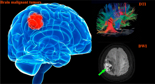
Previous studies showed a high diagnostic value of diffusion-weighted imaging (DWI) and diffusion tensor imaging (DTI) in differentiation among glioblastomas, primary cerebral lymphomas (PCLs), and solitary brain metastases, whereas other studies reported a low or no diagnostic value of DWI and DTI in differentiation among the three types of brain malignant tumors. In order to enhance the strength of evidence, meta-analysis was conducted to summarize results of studies evaluating the diagnostic values of DWI or DTI in differentiation among the three types of brain malignant tumors. Articles evaluating the diagnostic values of DWI or DTI in differentiation among the three types of tumors and published before December 2019 were searched in databases (PubMed, Medline, Web of Science, EMBASE, and Google Scholar). A summary of sensitivity, specificity, positive likelihood ratios (PLR), negative likelihood ratios (NLR), and diagnostic odds ratio (DOR) were calculated from the true positive (TP), true negative (TN), false positive (FP), and false negative (FN) of each study using STATA 12.0 software and Meta-Disc Version 1.4. In addition, the summary receive-operating characteristic (SROC) curve was constructed. Ultimately, we included 19 diagnostic studies (including 735 glioblastomas patients, 31 PCLs patients, and 792 patients with solitary brain metastases). Regarding differentiation between glioblastomas and solitary brain metastases using DWI or DTI, the calculated pooled parameters were as follows: sensitivity, 0.84 [95% confidence interval (CI): 0.78-0.89]; specificity, 0.88 (95% CI: 0.83-0.92); PLR, 7.2 (95% CI: 4.6-11.3); NLR, 0.18 (95% CI: 0.12-0.27); and DOR, 41 (95% CI: 18-93). The analysis showed a significant heterogeneity (sensitivity, I2 = 91.31%, p < 0.01; specificity, I2 = 89.24%, p < 0.01). In conclusion, DWI and DTI showed a moderate diagnostic value for differentiating glioblastomas from solitary brain metastasis. Additionally, large-scale prospective studies are essential to explore differentiation between PCLs and solitary brain metastases using DWI or DTI.
中文翻译:

胶质母细胞瘤,原发性脑淋巴瘤和孤立性脑转移之间的区别使用弥散加权成像和弥散张量成像:符合PRISMA的荟萃分析。
先前的研究表明弥散加权成像(DWI)和弥散张量成像(DTI)在胶质母细胞瘤,原发性脑淋巴瘤(PCL)和孤立性脑转移瘤之间的鉴别诊断中具有很高的诊断价值,而其他研究则报告低或无诊断价值。 DWI和DTI在三种类型的脑恶性肿瘤之间的分化。为了增强证据的强度,进行了荟萃分析,总结了评价DWI或DTI在三种类型的脑恶性肿瘤之间的诊断价值的研究结果。在数据库(PubMed,Medline,Web of Science,EMBASE和Google Scholar)中检索了评估DWI或DTI在三种类型的肿瘤之间的鉴别诊断价值并于2019年12月之前发表的文章。敏感性,特异性,阳性似然比(PLR),阴性似然比(NLR)和诊断比值比(DOR)由以下各项的真实正值(TP),真实负值(TN),假阳性(FP)和假阴性(FN)计算得出每项研究都使用STATA 12.0软件和Meta-Disc版本1.4。此外,绘制了汇总接收操作特性(SROC)曲线。最终,我们纳入了19项诊断研究(包括735例胶质母细胞瘤患者,31例PCL患者和792例患有孤立性脑转移的患者)。关于使用DWI或DTI区分胶质母细胞瘤和孤立性脑转移瘤,计算出的合并参数如下:敏感性,0.84 [95%置信区间(CI):0.78-0.89];特异性0.88(95%CI:0.83-0.92); PLR,7.2(95%CI:4.6-11.3);NLR:0.18(95%CI:0.12-0.27);DOR为41(95%CI:18-93)。分析显示出显着的异质性(灵敏度,I2 = 91.31%,p <0.01;特异性,I2 = 89.24%,p <0.01)。总之,DWI和DTI在区分胶质母细胞瘤和孤立性脑转移方面显示出中等诊断价值。此外,大规模前瞻性研究对于探索使用DWI或DTI的PCL和孤立性脑转移之间的区别至关重要。
更新日期:2020-01-10
中文翻译:
胶质母细胞瘤,原发性脑淋巴瘤和孤立性脑转移之间的区别使用弥散加权成像和弥散张量成像:符合PRISMA的荟萃分析。
先前的研究表明弥散加权成像(DWI)和弥散张量成像(DTI)在胶质母细胞瘤,原发性脑淋巴瘤(PCL)和孤立性脑转移瘤之间的鉴别诊断中具有很高的诊断价值,而其他研究则报告低或无诊断价值。 DWI和DTI在三种类型的脑恶性肿瘤之间的分化。为了增强证据的强度,进行了荟萃分析,总结了评价DWI或DTI在三种类型的脑恶性肿瘤之间的诊断价值的研究结果。在数据库(PubMed,Medline,Web of Science,EMBASE和Google Scholar)中检索了评估DWI或DTI在三种类型的肿瘤之间的鉴别诊断价值并于2019年12月之前发表的文章。敏感性,特异性,阳性似然比(PLR),阴性似然比(NLR)和诊断比值比(DOR)由以下各项的真实正值(TP),真实负值(TN),假阳性(FP)和假阴性(FN)计算得出每项研究都使用STATA 12.0软件和Meta-Disc版本1.4。此外,绘制了汇总接收操作特性(SROC)曲线。最终,我们纳入了19项诊断研究(包括735例胶质母细胞瘤患者,31例PCL患者和792例患有孤立性脑转移的患者)。关于使用DWI或DTI区分胶质母细胞瘤和孤立性脑转移瘤,计算出的合并参数如下:敏感性,0.84 [95%置信区间(CI):0.78-0.89];特异性0.88(95%CI:0.83-0.92); PLR,7.2(95%CI:4.6-11.3);NLR:0.18(95%CI:0.12-0.27);DOR为41(95%CI:18-93)。分析显示出显着的异质性(灵敏度,I2 = 91.31%,p <0.01;特异性,I2 = 89.24%,p <0.01)。总之,DWI和DTI在区分胶质母细胞瘤和孤立性脑转移方面显示出中等诊断价值。此外,大规模前瞻性研究对于探索使用DWI或DTI的PCL和孤立性脑转移之间的区别至关重要。


 京公网安备 11010802027423号