当前位置:
X-MOL 学术
›
J. Biophotonics
›
论文详情
Our official English website, www.x-mol.net, welcomes your
feedback! (Note: you will need to create a separate account there.)
Liver tissue classification of en face images by fractal dimension-based support vector machine.
Journal of Biophotonics ( IF 2.0 ) Pub Date : 2020-01-16 , DOI: 10.1002/jbio.201960154 Yue Zhu 1 , Wanrong Gao 1 , Zhenyan Guo 1 , Yawen Zhou 1 , Yuan Zhou 2
Journal of Biophotonics ( IF 2.0 ) Pub Date : 2020-01-16 , DOI: 10.1002/jbio.201960154 Yue Zhu 1 , Wanrong Gao 1 , Zhenyan Guo 1 , Yawen Zhou 1 , Yuan Zhou 2
Affiliation
|
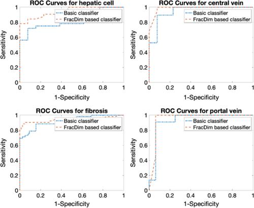
Full‐field optical coherence tomography (FF‐OCT) has been reported with its label‐free subcellular imaging performance. To realize quantitive cancer detection, the support vector machine model of classifying normal and cancerous human liver tissue is proposed with en face tomographic images. Twenty samples (10 normal and 10 cancerous) were operated from humans and composed of 285 en face tomographic images. Six histogram features and one proposed fractal dimension parameter that reveal the refractive index inhomogeneities of tissue were extracted and made up the training set. The other different 16 samples (8 normal and 8 cancerous) were imaged (190 images) and employed as the test set with the same features. First, a subcellular‐resolution tomographic image library for four histopathological areas in liver tissue was established. Second, the area under the receiver operating characteristics of 0.9378, 0.9858, 0.9391, 0.9517 for prediction of the cancerous hepatic cell, central vein, fibrosis, and portal vein were measured with the test set. The results indicate that the proposed classifier from FF‐OCT images shows promise as a label‐free assessment of quantified tumor detection, suggesting the fractal dimension‐based classifier could aid clinicians in detecting tumor boundaries for resection in surgery in the future.
中文翻译:

基于分形维的支持向量机对人脸图像进行肝组织分类。
据报道全场光学相干断层扫描(FF-OCT)具有无标记的亚细胞成像性能。为了实现定量癌症检测,提出了基于人脸断层图像对正常和癌性人类肝组织进行分类的支持向量机模型。从人类操作了20个样本(10个正常样本和10个癌样本),这些样本由285张全脸断层扫描图像组成。提取了六个直方图特征和一个建议的分形维参数,这些参数揭示了组织的折射率不均匀性,并组成了训练集。对其他16个样品(8个正常样品和8个癌样品)成像(190张图像)并用作具有相同特征的测试集。首先,建立了肝组织中四个组织病理学区域的亚细胞分辨率断层图像库。第二,用测试装置测量了接收器操作特性下的面积0.9378、0.9858、0.9391、0.9517,以预测癌细胞的肝细胞,中心静脉,纤维化和门静脉。结果表明,从FF-OCT图像中提出的分类器显示出有望作为无标记的量化肿瘤检测评估结果,表明基于分形维数的分类器可以帮助临床医生在未来的外科手术中检测肿瘤边界。
更新日期:2020-01-16
中文翻译:
基于分形维的支持向量机对人脸图像进行肝组织分类。
据报道全场光学相干断层扫描(FF-OCT)具有无标记的亚细胞成像性能。为了实现定量癌症检测,提出了基于人脸断层图像对正常和癌性人类肝组织进行分类的支持向量机模型。从人类操作了20个样本(10个正常样本和10个癌样本),这些样本由285张全脸断层扫描图像组成。提取了六个直方图特征和一个建议的分形维参数,这些参数揭示了组织的折射率不均匀性,并组成了训练集。对其他16个样品(8个正常样品和8个癌样品)成像(190张图像)并用作具有相同特征的测试集。首先,建立了肝组织中四个组织病理学区域的亚细胞分辨率断层图像库。第二,用测试装置测量了接收器操作特性下的面积0.9378、0.9858、0.9391、0.9517,以预测癌细胞的肝细胞,中心静脉,纤维化和门静脉。结果表明,从FF-OCT图像中提出的分类器显示出有望作为无标记的量化肿瘤检测评估结果,表明基于分形维数的分类器可以帮助临床医生在未来的外科手术中检测肿瘤边界。


 京公网安备 11010802027423号