当前位置:
X-MOL 学术
›
Neuroscience
›
论文详情
Our official English website, www.x-mol.net, welcomes your
feedback! (Note: you will need to create a separate account there.)
Enkephalinergic Circuit Involved in Nociceptive Modulation in the Spinal Dorsal Horn.
Neuroscience ( IF 2.9 ) Pub Date : 2020-01-07 , DOI: 10.1016/j.neuroscience.2019.12.020 Yang Bai 1 , Meng-Ying Li 2 , Jiang-Bo Ma 3 , Jia-Ni Li 1 , Xiao-Yu Teng 4 , Ying-Biao Chen 5 , Jun-Bin Yin 1 , Jing Huang 1 , Jing Chen 1 , Ting Zhang 1 , Xin-Tong Qiu 1 , Tao Chen 1 , Hui Li 1 , Sheng-Xi Wu 6 , Ya-Nan Peng 7 , Xiang Li 8 , Zhen-Zhen Kou 1 , Yun-Qing Li 9
Neuroscience ( IF 2.9 ) Pub Date : 2020-01-07 , DOI: 10.1016/j.neuroscience.2019.12.020 Yang Bai 1 , Meng-Ying Li 2 , Jiang-Bo Ma 3 , Jia-Ni Li 1 , Xiao-Yu Teng 4 , Ying-Biao Chen 5 , Jun-Bin Yin 1 , Jing Huang 1 , Jing Chen 1 , Ting Zhang 1 , Xin-Tong Qiu 1 , Tao Chen 1 , Hui Li 1 , Sheng-Xi Wu 6 , Ya-Nan Peng 7 , Xiang Li 8 , Zhen-Zhen Kou 1 , Yun-Qing Li 9
Affiliation
|
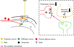
Enkephalin (ENK) has been implicated in pain modulation within the spinal dorsal horn (SDH). Revealing the mechanisms underlying ENK analgesia entails the anatomical and functional knowledge of spinal ENK-ergic circuits. Herein, we combined morphological and electrophysiological studies to unravel local ENK-ergic circuitry within the SDH. First, the distribution pattern of spinal ENK-ergic neurons was observed in adult preproenkephalin (PPE)-GFP knock-in mice. Next, the retrograde tracer tetramethylrhodamine (TMR) or horseradish peroxidase (HRP) was injected into the parabrachial nucleus (PBN) in PPE-GFP mice. Immunofluorescent staining showed I-isolectin B4 (IB4) labeled non-peptidergic afferents were in close apposition to TMR-labeled PBN-projecting neurons within lamina I as well as PPE-immunoreactivity (-ir) neurons within lamina II. Some TMR-labeled neurons were simultaneously in close association with both IB4 and PPE-ir terminals. Synaptic connections of these components were further confirmed by electron microscopy. Finally, TMR was injected into the PBN in adult C57BL/6 mice. Whole-cell patch recordings showed that δ-opioid receptor (DOR) agonist, [D-Pen2,5]-enkephalin (DPDPE, 1 µM), significantly reduced the frequency of miniature excitatory postsynaptic current (mEPSC) and decreased the activity of TMR-labeled neurons. In conclusion, spinal ENKergic neurons receive direct excitatory inputs from primary afferents, which might be directly recruited to release ENK under the condition of noxious stimuli; ENK could inhibit the glutamatergic transmission towards projecting neurons via presynaptic and postsynaptic DORs. These morphological and functional evidence may explain the mechanisms underlying the analgesic effects exerted by ENK within the SDH.
中文翻译:

脑脊髓回路参与脊髓背角的伤害性调节。
脑啡肽(ENK)与脊髓背角(SDH)内的疼痛调节有关。揭示ENK镇痛的基本机制需要脊柱ENK能量回路的解剖学和功能知识。在这里,我们结合了形态学和电生理学研究,以揭示SDH中的局部ENK能电路。首先,在成年前脑啡肽原(PPE)-GFP敲入小鼠中观察到脊髓ENK能神经元的分布模式。接下来,将逆行示踪剂四甲基罗丹明(TMR)或辣根过氧化物酶(HRP)注射到PPE-GFP小鼠的臂旁核(PBN)中。免疫荧光染色显示,I-异凝集素B4(IB4)标记的非肽类传入神经与层I内TMR标记的PBN投射神经元以及层II内的PPE免疫反应性(-ir)神经元紧密相关。一些TMR标记的神经元同时与IB4和PPE-ir末端密切相关。这些成分的突触连接通过电子显微镜进一步证实。最后,将TMR注射到成年C57BL / 6小鼠的PBN中。全细胞膜片记录表明,δ-阿片受体(DOR)激动剂[D-Pen2,5]-脑啡肽(DPDPE,1 µM)显着降低了小型兴奋性突触后电流(mEPSC)的频率并降低了TMR的活性标记的神经元。综上所述,脊神经ENKergic神经元从原发神经传入直接兴奋性输入,在有毒刺激的情况下,可直接募集以释放ENK。ENK可以通过突触前和突触后DOR抑制谷氨酸能向投射神经元的传递。
更新日期:2020-01-07
中文翻译:
脑脊髓回路参与脊髓背角的伤害性调节。
脑啡肽(ENK)与脊髓背角(SDH)内的疼痛调节有关。揭示ENK镇痛的基本机制需要脊柱ENK能量回路的解剖学和功能知识。在这里,我们结合了形态学和电生理学研究,以揭示SDH中的局部ENK能电路。首先,在成年前脑啡肽原(PPE)-GFP敲入小鼠中观察到脊髓ENK能神经元的分布模式。接下来,将逆行示踪剂四甲基罗丹明(TMR)或辣根过氧化物酶(HRP)注射到PPE-GFP小鼠的臂旁核(PBN)中。免疫荧光染色显示,I-异凝集素B4(IB4)标记的非肽类传入神经与层I内TMR标记的PBN投射神经元以及层II内的PPE免疫反应性(-ir)神经元紧密相关。一些TMR标记的神经元同时与IB4和PPE-ir末端密切相关。这些成分的突触连接通过电子显微镜进一步证实。最后,将TMR注射到成年C57BL / 6小鼠的PBN中。全细胞膜片记录表明,δ-阿片受体(DOR)激动剂[D-Pen2,5]-脑啡肽(DPDPE,1 µM)显着降低了小型兴奋性突触后电流(mEPSC)的频率并降低了TMR的活性标记的神经元。综上所述,脊神经ENKergic神经元从原发神经传入直接兴奋性输入,在有毒刺激的情况下,可直接募集以释放ENK。ENK可以通过突触前和突触后DOR抑制谷氨酸能向投射神经元的传递。


 京公网安备 11010802027423号