Nature Methods ( IF 36.1 ) Pub Date : 2020-01-06 , DOI: 10.1038/s41592-019-0676-4 Yongdeng Zhang 1 , Lena K Schroeder 1 , Mark D Lessard 1 , Phylicia Kidd 1 , Jeeyun Chung 1, 2, 3 , Yuanbin Song 4 , Lorena Benedetti 1, 2, 3 , Yiming Li 5 , Jonas Ries 5 , Jonathan B Grimm 6 , Luke D Lavis 6 , Pietro De Camilli 1, 2, 3, 7 , James E Rothman 1, 8 , David Baddeley 1, 8, 9 , Joerg Bewersdorf 1, 7, 8, 10
|
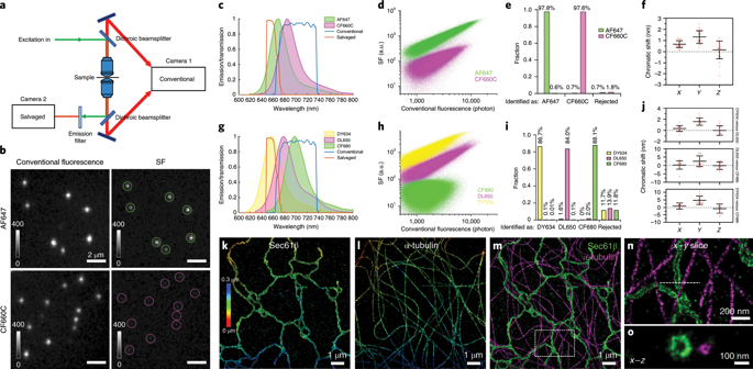
Combining the molecular specificity of fluorescent probes with three-dimensional imaging at nanoscale resolution is critical for investigating the spatial organization and interactions of cellular organelles and protein complexes. We present a 4Pi single-molecule switching super-resolution microscope that enables ratiometric multicolor imaging of mammalian cells at 5–10-nm localization precision in three dimensions using ‘salvaged fluorescence’. Imaging two or three fluorophores simultaneously, we show fluorescence images that resolve the highly convoluted Golgi apparatus and the close contacts between the endoplasmic reticulum and the plasma membrane, structures that have traditionally been the imaging realm of electron microscopy. The salvaged fluorescence approach is equally applicable in most single-objective microscopes.
中文翻译:
多色三维回收荧光成像揭示的纳米级亚细胞结构
将荧光探针的分子特异性与纳米级分辨率的三维成像相结合对于研究细胞器和蛋白质复合物的空间组织和相互作用至关重要。我们展示了一种 4Pi 单分子切换超分辨率显微镜,该显微镜能够使用“回收荧光”以 5–10 nm 的三维定位精度对哺乳动物细胞进行比例多色成像。同时对两个或三个荧光团进行成像,我们显示的荧光图像解析了高度复杂的高尔基体以及内质网和质膜之间的紧密接触,这些结构在传统上一直是电子显微镜的成像领域。回收荧光方法同样适用于大多数单物镜显微镜。


 京公网安备 11010802027423号