Nature Medicine ( IF 58.7 ) Pub Date : 2020-01-06 , DOI: 10.1038/s41591-019-0715-9 Todd C Hollon 1 , Balaji Pandian 2 , Arjun R Adapa 2 , Esteban Urias 2 , Akshay V Save 3 , Siri Sahib S Khalsa 1 , Daniel G Eichberg 4 , Randy S D'Amico 5 , Zia U Farooq 6 , Spencer Lewis 2 , Petros D Petridis 3 , Tamara Marie 7 , Ashish H Shah 4 , Hugh J L Garton 1 , Cormac O Maher 1 , Jason A Heth 1 , Erin L McKean 1, 8 , Stephen E Sullivan 1 , Shawn L Hervey-Jumper 1, 9 , Parag G Patil 1 , B Gregory Thompson 1 , Oren Sagher 1 , Guy M McKhann 5 , Ricardo J Komotar 4 , Michael E Ivan 4 , Matija Snuderl 10 , Marc L Otten 5 , Timothy D Johnson 11 , Michael B Sisti 5 , Jeffrey N Bruce 5 , Karin M Muraszko 1 , Jay Trautman 6 , Christian W Freudiger 6 , Peter Canoll 12 , Honglak Lee 13 , Sandra Camelo-Piragua 14 , Daniel A Orringer 1, 15
|
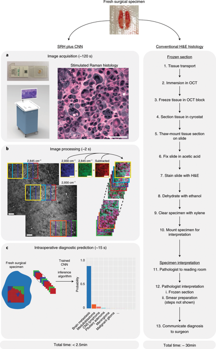
Intraoperative diagnosis is essential for providing safe and effective care during cancer surgery1. The existing workflow for intraoperative diagnosis based on hematoxylin and eosin staining of processed tissue is time, resource and labor intensive2,3. Moreover, interpretation of intraoperative histologic images is dependent on a contracting, unevenly distributed, pathology workforce4. In the present study, we report a parallel workflow that combines stimulated Raman histology (SRH)5,6,7, a label-free optical imaging method and deep convolutional neural networks (CNNs) to predict diagnosis at the bedside in near real-time in an automated fashion. Specifically, our CNNs, trained on over 2.5 million SRH images, predict brain tumor diagnosis in the operating room in under 150 s, an order of magnitude faster than conventional techniques (for example, 20–30 min)2. In a multicenter, prospective clinical trial (n = 278), we demonstrated that CNN-based diagnosis of SRH images was noninferior to pathologist-based interpretation of conventional histologic images (overall accuracy, 94.6% versus 93.9%). Our CNNs learned a hierarchy of recognizable histologic feature representations to classify the major histopathologic classes of brain tumors. In addition, we implemented a semantic segmentation method to identify tumor-infiltrated diagnostic regions within SRH images. These results demonstrate how intraoperative cancer diagnosis can be streamlined, creating a complementary pathway for tissue diagnosis that is independent of a traditional pathology laboratory.
中文翻译:
使用受激拉曼组织学和深度神经网络进行近实时术中脑肿瘤诊断
术中诊断对于在癌症手术期间提供安全有效的护理至关重要1 。基于处理组织的苏木精和伊红染色的现有术中诊断工作流程是时间、资源和劳动力密集型的2,3 。此外,术中组织学图像的解释取决于收缩的、分布不均匀的病理学人员4 。在本研究中,我们报告了一种并行工作流程,该工作流程结合了受激拉曼组织学 (SRH) 5,6,7 、一种无标记光学成像方法和深度卷积神经网络 (CNN),可近乎实时地预测床边诊断以自动化的方式。具体来说,我们的 CNN 经过超过 250 万张 SRH 图像的训练,可在 150 秒内预测手术室中的脑肿瘤诊断,比传统技术(例如 20-30 分钟)快一个数量级2 。在一项多中心、前瞻性临床试验 ( n = 278) 中,我们证明基于 CNN 的 SRH 图像诊断并不劣于基于病理学家的传统组织学图像解释(总体准确率,94.6% 与 93.9%)。我们的 CNN 学习了可识别的组织学特征表示的层次结构,以对脑肿瘤的主要组织病理学类别进行分类。此外,我们还实施了语义分割方法来识别 SRH 图像中肿瘤浸润的诊断区域。这些结果证明了如何简化术中癌症诊断,为独立于传统病理实验室的组织诊断创建补充途径。


 京公网安备 11010802027423号