Journal of Neuroradiology ( IF 3.0 ) Pub Date : 2019-09-26 , DOI: 10.1016/j.neurad.2019.08.004 Y Abe 1 , I Yuki 2 , K Otani 3 , T Shoji 1 , T Ishibashi 2 , Y Murayama 2
|
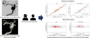
Background and purpose
Precise vessel measurement plays a major role in size selection of stents used for the treatment of intracranial aneurysms and became even more critical after the introduction of flow diverter stents. We assessed agreement between intracranial vessel diameters of aneurysm patients measured on 2D digital subtraction (2D DSA) and 3D volume rendering digital subtraction angiography (3D DSA) images using an automatic windowing algorithm.
Materials and methods
Ten patients with intracranial aneurysms were enrolled and 120 measurement points were selected on both 2D and 3D DSA images acquired by a biplane angiographic system. Automatic windowing was applied to the 3D DSA images. Inter-method agreement of vessel measurements on 2D and 3D DSA images was assessed by Bland Altman plots and intraclass correlation coefficients (ICC). Inter- and intra-rater agreement of measurements on 3D DSA images were assessed by ICCs.
Results
The mean differences between measurements on 2D and 3D DSA images were 0.14 mm for the ICA, and 0.18 mm for the ACA and MCA, which is about the size of one 3D DSA image voxel. For ICA measurements, inter-method, inter-rater and intra-rater agreements were good or excellent (consistency and absolute ICC ≥ 0.95). For ACA and MCA measurements, the inter-method, inter-rater and intra-rater agreements were also good or excellent (consistency ICC = 0.94, 0.89 and 0.93, absolute ICC = 0.83, 0.84 and 0.85 respectively).
Conclusions
Vessel diameters may be measured on 3D DSA images with sufficient reliability for clinical use when applying an automatic windowing algorithm.
中文翻译:
使用自动加窗算法在 2D 和 3D 数字减影血管造影术上测量的颅内血管直径的一致性
背景和目的
精确的血管测量在用于治疗颅内动脉瘤的支架尺寸选择中起着重要作用,并且在引入分流器支架后变得更加重要。我们使用自动加窗算法评估了在 2D 数字减影 (2D DSA) 和 3D 体积渲染数字减影血管造影 (3D DSA) 图像上测量的动脉瘤患者颅内血管直径之间的一致性。
材料和方法
招募了 10 名颅内动脉瘤患者,并在双平面血管造影系统采集的 2D 和 3D DSA 图像上选择了 120 个测量点。自动开窗应用于 3D DSA 图像。血管测量在 2D 和 3D DSA 图像上的方法间一致性通过 Bland Altman 图和组内相关系数 (ICC) 进行评估。ICC 评估了 3D DSA 图像测量的内部和内部一致性。
结果
ICA 的2D 和 3D DSA 图像测量值之间的平均差异为 0.14 mm, ACA 和 MCA 的测量值之间的平均差异为 0.18 mm,大约是一个 3D DSA 图像体素的大小。对于 ICA 测量,方法间、评估者间和评估者内的一致性是好的或优秀的(一致性和绝对 ICC ≥ 0.95)。对于 ACA 和 MCA 测量,方法间、评估者间和评估者内部的一致性也很好或非常好(一致性 ICC = 0.94、0.89 和 0.93,绝对 ICC 分别 = 0.83、0.84 和 0.85)。
结论
当应用自动加窗算法时,血管直径可以在 3D DSA 图像上测量,具有足够的临床使用可靠性。


 京公网安备 11010802027423号