Journal of Neuroradiology ( IF 3.0 ) Pub Date : 2019-08-08 , DOI: 10.1016/j.neurad.2019.06.005 Maxence Serru 1 , Bénédicte Marechal 2 , Tobias Kober 2 , Leo Ribier 1 , Catherine Sembely Taveau 1 , Dominique Sirinelli 3 , Jean-Philippe Cottier 4 , Baptiste Morel 3
|
Background and purpose
It can be challenging to depict brain volume abnormalities in the pediatric population on magnetic resonance imaging (MRI). The aim of the study was to evaluate the inter-radiologist reliability in brain MRI interpretation, including brain volume assessment and the efficiency of an automated brain segmentation.
Materials and methods
We performed a single-center prospective study including 44 patients aged six months to five years recruited from the University Hospital, having a 1.5 T brain MRI using a MP2RAGE sequence. All MRI were randomly and blindly reviewed by one junior and two senior pediatric radiologists. Inter-observer agreements were assessed using Fleiss’ kappa coefficient. Brain volumetry (total intracranial volume (TIV), brain parenchyma, and cerebrospinal fluid volumes) was estimated using the MorphoBox prototype. Clinical head circumference (HC) and z scores were reported. A Pearson correlation coefficient was calculated between brain volumes with HC.
Results
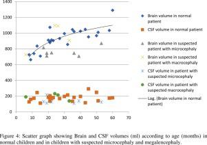
Twenty-four brain MRI examinations were normal and twenty were pathological. Brain volume abnormalities were poorly detected by junior and senior radiologists: sensitivities 16.67% [confidence interval 4.7–44.8], 33.33% [13–60] and 30.7% [12–58] and specificities 93.75% [79–98], 84.38% [68–93] and 77% [60–88], respectively. Brain volume apart, interobserver kappa coefficients were 0.93 between junior and seniors as well as between seniors. Brain volumes were significantly correlated with HC (P < 0.0001). In patients with normal MRI, brain parenchyma volumes increased regularly with age. Low brain volume was easier to identify with automated quantification.
Conclusion
Brain volume was poorly appreciated by radiologists. The fully automated brain segmentation used can provide quantitative data to better diagnose, describe, and follow-up brain volume abnormalities.
中文翻译:
使用基于 MP2RAGE 的自动 MRI 脑分割提高儿童期脑容量异常的诊断准确性
背景和目的
在磁共振成像 (MRI) 上描绘儿科人群的脑容量异常可能具有挑战性。该研究的目的是评估脑 MRI 解释中放射科医生间的可靠性,包括脑容量评估和自动脑分割的效率。
材料和方法
我们进行了一项单中心前瞻性研究,包括从大学医院招募的 44 名 6 个月至 5 岁的患者,他们 使用 MP2RAGE 序列进行了 1.5 T 的脑部 MRI。所有 MRI 均由一名初级和两名高级儿科放射科医生随机盲审。使用 Fleiss 的 kappa 系数评估观察者间的协议。使用 MorphoBox 原型估计脑容量(总颅内容量 (TIV)、脑实质和脑脊液容量)。报告了临床头围 (HC) 和 z 评分。计算脑容量与 HC 之间的 Pearson 相关系数。
结果
24 例脑部 MRI 检查正常,20 例为病理检查。初级和高级放射科医生很难检测到脑容量异常:敏感性为 16.67% [置信区间 4.7-44.8]、33.33% [13-60] 和 30.7% [12-58],特异性为 93.75% [79-98]、84.38% [68-93] 和 77% [60-88],分别。除了脑容量,初级和高级之间以及高级之间的观察者间 kappa 系数为 0.93。脑容量与 HC 显着相关(P < 0.0001)。在 MRI 正常的患者中,脑实质体积随着年龄的增长而有规律地增加。通过自动量化更容易识别低脑容量。
结论
放射科医生对脑容量的评价不高。使用的全自动大脑分割可以提供定量数据,以更好地诊断、描述和跟踪脑容量异常。


 京公网安备 11010802027423号