Journal of Neuroradiology ( IF 3.0 ) Pub Date : 2019-06-20 , DOI: 10.1016/j.neurad.2019.05.006 Yukitomo Ishi 1 , Shigeru Yamaguchi 1 , Michiharu Yoshida 1 , Hiroaki Motegi 1 , Hiroyuki Kobayashi 1 , Shunsuke Terasaka 1 , Kiyohiro Houkin 1
|
Background and purpose
Most individuals with optic pathway/hypothalamic pilocytic astrocytoma (OPHPA) harbor either the BRAF V600E mutation or KIAA1549-BRAF fusion (K-B). This study aimed to investigate the imaging characteristics of OPHPA in relation to BRAF alteration status.
Materials and methods
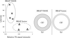
Seven cases of OPHPA harboring either the BRAF V600E mutation or K-B fusion were included in the study. Preoperative magnetic resonance imaging (MRI) was assessed for degree of T2 hyperintensity on T2-weighted images (T2WI) and the ratio of nonenhancing T2 or fluid-attenuated inversion recovery (FLAIR) hyperintense area to the contrast enhanced area (CE) on gadolinium-enhanced-T1 weighted images (T2/FLAIR-CE mismatch). The T2 signal intensity was normalized to cerebrospinal fluid (T2/CSF) for both the V600E and K-B group and compared. T2/FLAIR-CE mismatch was assessed by calculating the proportion of the tumor volume of nonenhancing high T2 signal intensity to the whole lesion (nonenhancing and enhancing components).
Results
Four and three cases of OPHPA harboring the BRAF V600E mutation and K-B, respectively, were analyzed. The T2/CSF value was higher in the K-B group than in the V600E group. Moreover, the V600E group had a larger T2/FLAIR-CE mismatch than the K-B group.
Conclusions
The BRAF alteration status in individuals with OPHPA was associated with preoperative MRI by focusing on T2 signal intensity and T2/FLAIR-CE mismatch. The BRAF V600E mutation was associated with a lower T2/CSF value and larger T2/FLAIR-CE mismatch, whereas K-B fusion was associated with a higher T2/CSF value and smaller T2/FLAIR-CE mismatch.
中文翻译:
视通路/下丘脑毛细胞星形细胞瘤患者磁共振成像特征与 BRAF 改变状态的相关性
背景和目的
大多数患有视通路/下丘脑毛细胞星形细胞瘤 (OPHPA) 的个体携带BRAF V600E 突变或KIAA1549-BRAF融合 (KB)。本研究旨在探讨 OPHPA 与BRAF改变状态相关的影像学特征。
材料和方法
该研究包括 7 例携带BRAF V600E 突变或 KB 融合的 OPHPA 病例。术前磁共振成像 (MRI) 评估 T2 加权图像 (T2WI) 上的 T2 高信号程度以及非增强 T2 或流体衰减反转恢复 (FLAIR) 高信号区域与钆对比增强区域 (CE) 的比率-增强 T1 加权图像(T2/FLAIR-CE 不匹配)。将 V600E 和 KB 组的 T2 信号强度标准化为脑脊液 (T2/CSF) 并进行比较。通过计算非增强高 T2 信号强度的肿瘤体积占整个病灶(非增强和增强成分)的比例来评估 T2/FLAIR-CE 不匹配。
结果
分别分析了 4 例和 3 例携带 BRAF V600E 突变和 KB 的 OPHPA。KB 组的 T2/CSF 值高于 V600E 组。此外,V600E 组的 T2/FLAIR-CE 失配比 KB 组更大。
结论
通过关注 T2 信号强度和 T2/FLAIR-CE 不匹配,OPHPA 患者的 BRAF 改变状态与术前 MRI 相关。BRAF V600E 突变与较低的 T2/CSF 值和较大的 T2/FLAIR-CE 错配相关,而 KB 融合与较高的 T2/CSF 值和较小的 T2/FLAIR-CE 错配相关。


 京公网安备 11010802027423号