Our official English website, www.x-mol.net, welcomes your
feedback! (Note: you will need to create a separate account there.)
High-Dimensional Analysis Delineates Myeloid and Lymphoid Compartment Remodeling during Successful Immune-Checkpoint Cancer Therapy
Cell ( IF 45.5 ) Pub Date : 2018-11-15 00:00:00 , DOI: 10.1016/j.cell.2018.11.003 Matthew M. Gubin , Ekaterina Esaulova , Jeffrey P. Ward , Olga N. Malkova , Daniele Runci , Pamela Wong , Takuro Noguchi , Cora D. Arthur , Wei Meng , Elise Alspach , Ruan F.V. Medrano , Catrina Fronick , Michael Fehlings , Evan W. Newell , Robert S. Fulton , Kathleen C.F. Sheehan , Stephen T. Oh , Robert D. Schreiber , Maxim N. Artyomov
中文翻译:

高维分析描述了成功的免疫检查点癌症治疗过程中的髓样和淋巴隔室重塑
更新日期:2018-11-15
Cell ( IF 45.5 ) Pub Date : 2018-11-15 00:00:00 , DOI: 10.1016/j.cell.2018.11.003 Matthew M. Gubin , Ekaterina Esaulova , Jeffrey P. Ward , Olga N. Malkova , Daniele Runci , Pamela Wong , Takuro Noguchi , Cora D. Arthur , Wei Meng , Elise Alspach , Ruan F.V. Medrano , Catrina Fronick , Michael Fehlings , Evan W. Newell , Robert S. Fulton , Kathleen C.F. Sheehan , Stephen T. Oh , Robert D. Schreiber , Maxim N. Artyomov
|
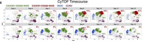
(Cell175, 1014–1030.e1–e19; November 1, 2018) Due to an error in the preparation of the figure, the labels for CX3CR1−CD206+iNOS− and CX3CR1+CD206+iNOS− cells were inverted in Figure 6E. This has been corrected in the online version of the article, and the correct version of the figure panel is reproduced below.Figure 6ERestructuring of Intratumoral Monocytes/Macrophages Revealed by Longitudinal Analyses (original)View Large ImageFigure ViewerDownload Hi-res imageDownload (PPT)
中文翻译:
高维分析描述了成功的免疫检查点癌症治疗过程中的髓样和淋巴隔室重塑
(Cell175,1014-1030.e1-e19; 2018年11月1日)由于制图错误,CX3CR1-CD206 + iNOS-和CX3CR1 + CD206 + iNOS-细胞的标签在图6E中被颠倒了。此问题已在文章的在线版本中得到纠正,并且图面板的正确版本如下所示。图6纵向分析揭示的肿瘤内单核细胞/巨噬细胞的结构(原始)查看大图图ViewerDownload高分辨率imageDownload(PPT)


 京公网安备 11010802027423号