Our official English website, www.x-mol.net, welcomes your
feedback! (Note: you will need to create a separate account there.)
Reflection phase microscopy using spatio-temporal coherence of light
Optica ( IF 8.4 ) Pub Date : 2018-11-15 , DOI: 10.1364/optica.5.001468 Youngwoon Choi 1, 2 , Poorya Hosseini 3 , Jeon Woong Kang 3 , Sungsam Kang 3 , Taeseok Daniel Yang 1 , Min Gyu Hyeon 4 , Beop-Min Kim 1, 2, 4 , Peter T C So 3, 5 , Zahid Yaqoob 3
Optica ( IF 8.4 ) Pub Date : 2018-11-15 , DOI: 10.1364/optica.5.001468 Youngwoon Choi 1, 2 , Poorya Hosseini 3 , Jeon Woong Kang 3 , Sungsam Kang 3 , Taeseok Daniel Yang 1 , Min Gyu Hyeon 4 , Beop-Min Kim 1, 2, 4 , Peter T C So 3, 5 , Zahid Yaqoob 3
Affiliation
|
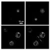
Many disease states are associated with cellular biomechanical changes as markers. Label-free phase microscopes are used to quantify thermally driven interface fluctuations, which allow the deduction of important cellular rheological properties. Here, the spatio-temporal coherence of light was used to implement a high-speed reflection phase microscope with superior depth selectivity and higher phase sensitivity. Nanometric scale motion of cytoplasmic structures can be visualized with fine details and three-dimensional resolution. Specifically, the spontaneous fluctuation occurring on the nuclear membrane of a living cell was observed at video rate. By converting the reflection phase into displacement, the sensitivity in quantifying nuclear membrane fluctuation was found to be about one nanometer. A reflection phase microscope can potentially elucidate biomechanical mechanisms of pathological and physiological processes.
中文翻译:

利用光的时空相干性的反射相位显微镜
许多疾病状态都与作为标记的细胞生物力学变化相关。无标记相显微镜用于量化热驱动的界面波动,从而可以推断出重要的细胞流变特性。在这里,利用光的时空相干性来实现具有优异的深度选择性和更高的相位灵敏度的高速反射相位显微镜。细胞质结构的纳米级运动可以以精细的细节和三维分辨率可视化。具体来说,以视频速率观察活细胞核膜上发生的自发波动。通过将反射相位转换为位移,发现量化核膜波动的灵敏度约为一纳米。反射相位显微镜可以潜在地阐明病理和生理过程的生物力学机制。
更新日期:2018-11-21
中文翻译:
利用光的时空相干性的反射相位显微镜
许多疾病状态都与作为标记的细胞生物力学变化相关。无标记相显微镜用于量化热驱动的界面波动,从而可以推断出重要的细胞流变特性。在这里,利用光的时空相干性来实现具有优异的深度选择性和更高的相位灵敏度的高速反射相位显微镜。细胞质结构的纳米级运动可以以精细的细节和三维分辨率可视化。具体来说,以视频速率观察活细胞核膜上发生的自发波动。通过将反射相位转换为位移,发现量化核膜波动的灵敏度约为一纳米。反射相位显微镜可以潜在地阐明病理和生理过程的生物力学机制。


 京公网安备 11010802027423号