当前位置:
X-MOL 学术
›
J. Phys. Chem. Lett.
›
论文详情
Our official English website, www.x-mol.net, welcomes your
feedback! (Note: you will need to create a separate account there.)
SERS and Cryo-EM Directly Reveal Different Liposome Structures during Interaction with Gold Nanoparticles
The Journal of Physical Chemistry Letters ( IF 4.8 ) Pub Date : 2018-11-13 00:00:00 , DOI: 10.1021/acs.jpclett.8b03191 Vesna Živanović 1, 2 , Zdravko Kochovski 3 , Christoph Arenz 1, 2 , Yan Lu 3, 4 , Janina Kneipp 1, 2
The Journal of Physical Chemistry Letters ( IF 4.8 ) Pub Date : 2018-11-13 00:00:00 , DOI: 10.1021/acs.jpclett.8b03191 Vesna Živanović 1, 2 , Zdravko Kochovski 3 , Christoph Arenz 1, 2 , Yan Lu 3, 4 , Janina Kneipp 1, 2
Affiliation
|
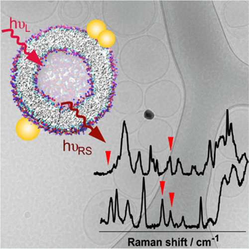
The combination of gold nanoparticles with liposomes is important for nano- and biotechnology. Here, we present direct, label-free characterization of liposome structure and composition at the site of its interaction with citrate-stabilized gold nanoparticles by surface-enhanced Raman scattering (SERS) and cryogenic electron microscopy (cryo-EM). Evidenced by the vibrational spectra and cryo-EM, the gold nanoparticles destroy the bilayer structure of interacting liposomes in the presence of a high amount of citrate, while at lower citrate concentration the nanoparticles interact with the surface of the intact liposomes. The spectra of phosphatidylcholine and phosphatidylcholine/sphingomyelin liposomes show that at the site of interaction the lipid chains are in the gel phase. The SERS spectra indicate that cholesterol has strong effects on the contacts of the vesicles with the nanoparticles. By combining cryo-EM and SERS, the structure and properties of lipid–nanoparticle composites could be tailored for the development of drug delivery systems.
中文翻译:

SERS和Cryo-EM在与金纳米粒子相互作用期间直接揭示不同的脂质体结构。
金纳米颗粒与脂质体的结合对于纳米技术和生物技术很重要。在这里,我们通过表面增强拉曼散射(SERS)和低温电子显微镜(cryo-EM),在脂质体与柠檬酸盐稳定的金纳米颗粒相互作用的位点上,提供脂质体结构和组成的直接,无标记的表征。通过振动光谱和冷冻EM证明,金纳米颗粒在存在大量柠檬酸盐的情况下破坏了相互作用脂质体的双层结构,而在柠檬酸盐浓度较低时,纳米颗粒与完整脂质体的表面相互作用。磷脂酰胆碱和磷脂酰胆碱/鞘磷脂脂质体的光谱表明,在相互作用位点,脂质链处于凝胶相。SERS光谱表明胆固醇对囊泡与纳米颗粒的接触具有强烈影响。通过结合cryo-EM和SERS,可以针对脂质-纳米颗粒复合材料的结构和性能进行定制,以开发药物输送系统。
更新日期:2018-11-13
中文翻译:
SERS和Cryo-EM在与金纳米粒子相互作用期间直接揭示不同的脂质体结构。
金纳米颗粒与脂质体的结合对于纳米技术和生物技术很重要。在这里,我们通过表面增强拉曼散射(SERS)和低温电子显微镜(cryo-EM),在脂质体与柠檬酸盐稳定的金纳米颗粒相互作用的位点上,提供脂质体结构和组成的直接,无标记的表征。通过振动光谱和冷冻EM证明,金纳米颗粒在存在大量柠檬酸盐的情况下破坏了相互作用脂质体的双层结构,而在柠檬酸盐浓度较低时,纳米颗粒与完整脂质体的表面相互作用。磷脂酰胆碱和磷脂酰胆碱/鞘磷脂脂质体的光谱表明,在相互作用位点,脂质链处于凝胶相。SERS光谱表明胆固醇对囊泡与纳米颗粒的接触具有强烈影响。通过结合cryo-EM和SERS,可以针对脂质-纳米颗粒复合材料的结构和性能进行定制,以开发药物输送系统。


 京公网安备 11010802027423号