当前位置:
X-MOL 学术
›
Faraday Discuss.
›
论文详情
Our official English website, www.x-mol.net, welcomes your
feedback! (Note: you will need to create a separate account there.)
The role of a plasmonic substrate on the enhancement and spatial resolution of tip-enhanced Raman scattering
Faraday Discussions ( IF 3.3 ) Pub Date : 2018-11-07 , DOI: 10.1039/c8fd00142a Mahfujur Rahaman 1, 2, 3, 4 , Alexander G. Milekhin 5, 6, 6, 7, 8 , Ashutosh Mukherjee 1, 2, 3, 4 , Ekaterina E. Rodyakina 5, 6, 6, 7, 8 , Alexander V. Latyshev 5, 6, 6, 7, 8 , Volodymyr M. Dzhagan 1, 2, 3, 4, 9 , Dietrich R. T. Zahn 1, 2, 3, 4
Faraday Discussions ( IF 3.3 ) Pub Date : 2018-11-07 , DOI: 10.1039/c8fd00142a Mahfujur Rahaman 1, 2, 3, 4 , Alexander G. Milekhin 5, 6, 6, 7, 8 , Ashutosh Mukherjee 1, 2, 3, 4 , Ekaterina E. Rodyakina 5, 6, 6, 7, 8 , Alexander V. Latyshev 5, 6, 6, 7, 8 , Volodymyr M. Dzhagan 1, 2, 3, 4, 9 , Dietrich R. T. Zahn 1, 2, 3, 4
Affiliation

|
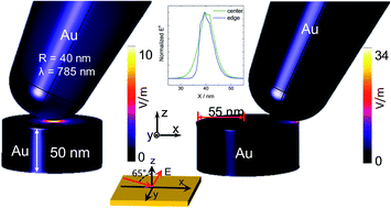
Since the first report in the early 2000s, there have been several experimental configurations that have demonstrated enhancement and spatial resolution of tip-enhanced Raman spectroscopy (TERS). The combination of a plasmonic substrate and a metallic tip is one suitable approach to achieve even higher enhancement and lateral resolution. In this contribution, we demonstrate TERS on a monolayer of MoS2 on an array of Au nanodisks. The Au nanodisks were prepared by electron beam writing. Thereafter, MoS2 was transferred onto the plasmonic substrate via the exfoliation technique. We witness an unprecedented enhancement and spatial resolution in the experiments. In the TERS image a ring-like shape is observed that matches the edges of the nanodisks. TERS enhancement at the edges is about 170 times stronger than at the center of the nanodisks. For a better understanding of the experimental results, finite element method (FEM) simulations were employed to simulate the TERS image of the MoS2/plasmonic heterostructure. Our calculations show a higher electric field concentration at the edges that exponentially decays to the center. Therefore, it reproduces the ring-like shape of the experimental image. Moreover, the calculations suggest a TERS enhancement of 135 at the edges compared to the center, which is in very good agreement with the experimental data. According to our calculations, the spatial resolution is also increased at the edges. For comparison, FEM simulations of a tip–flat metal substrate system (conventional gap-mode TERS) were carried out. The calculations confirmed a 110 times stronger enhancement at the edges of the nanodisks than that of conventional gap-mode TERS and explained the experimental maps. Our results provide not only a deeper understanding of the TERS mechanism of this heterostructure, but can also help in realizing highly efficient TERS experiments using similar systems.
中文翻译:

等离子体基质对尖端增强拉曼散射的增强和空间分辨率的作用
自2000年代初的第一份报告以来,已经有几种实验配置证明了尖端增强拉曼光谱(TERS)的增强和空间分辨率。等离子体衬底和金属尖端的组合是一种获得更高增强和横向分辨率的合适方法。在这项贡献中,我们证明了在Au纳米磁盘阵列上MoS 2单层上的TERS 。通过电子束写入制备金纳米盘。此后,的MoS 2转移到等离激元基底通过去角质技术。我们见证了实验中前所未有的增强和空间分辨率。在TERS图像中,观察到与纳米盘边缘匹配的环形形状。边缘的TERS增强比纳米磁盘的中心强170倍。为了更好地了解实验结果,采用了有限元方法(FEM)模拟来模拟MoS 2的TERS图像/等离子体异质结构。我们的计算结果表明,边缘处的电场集中度较高,并呈指数衰减至中心。因此,它再现了实验图像的环形形状。此外,计算结果表明,与中心相比,边缘的TERS增强了135,这与实验数据非常吻合。根据我们的计算,边缘处的空间分辨率也会提高。为了进行比较,对尖端-扁平金属基板系统(传统的间隙模式TERS)进行了有限元模拟。计算结果证实,纳米盘边缘的增强作用是传统间隙模式TERS的110倍,并解释了实验图。我们的结果不仅提供了对这种异质结构的TERS机制的更深入的了解,
更新日期:2019-05-29
中文翻译:

等离子体基质对尖端增强拉曼散射的增强和空间分辨率的作用
自2000年代初的第一份报告以来,已经有几种实验配置证明了尖端增强拉曼光谱(TERS)的增强和空间分辨率。等离子体衬底和金属尖端的组合是一种获得更高增强和横向分辨率的合适方法。在这项贡献中,我们证明了在Au纳米磁盘阵列上MoS 2单层上的TERS 。通过电子束写入制备金纳米盘。此后,的MoS 2转移到等离激元基底通过去角质技术。我们见证了实验中前所未有的增强和空间分辨率。在TERS图像中,观察到与纳米盘边缘匹配的环形形状。边缘的TERS增强比纳米磁盘的中心强170倍。为了更好地了解实验结果,采用了有限元方法(FEM)模拟来模拟MoS 2的TERS图像/等离子体异质结构。我们的计算结果表明,边缘处的电场集中度较高,并呈指数衰减至中心。因此,它再现了实验图像的环形形状。此外,计算结果表明,与中心相比,边缘的TERS增强了135,这与实验数据非常吻合。根据我们的计算,边缘处的空间分辨率也会提高。为了进行比较,对尖端-扁平金属基板系统(传统的间隙模式TERS)进行了有限元模拟。计算结果证实,纳米盘边缘的增强作用是传统间隙模式TERS的110倍,并解释了实验图。我们的结果不仅提供了对这种异质结构的TERS机制的更深入的了解,


 京公网安备 11010802027423号