Our official English website, www.x-mol.net, welcomes your
feedback! (Note: you will need to create a separate account there.)
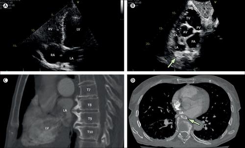
External left atrium compression by spinal osteophytes
The Lancet ( IF 98.4 ) Pub Date : 2018-10-20 , DOI: 10.1016/s0140-6736(18)32282-7 Hourmazd Haghbayan , Eric A Coomes , Asim N Cheema
The Lancet ( IF 98.4 ) Pub Date : 2018-10-20 , DOI: 10.1016/s0140-6736(18)32282-7 Hourmazd Haghbayan , Eric A Coomes , Asim N Cheema
|
A previously healthy 76-year-old woman was assessed preoperatively prior to hip arthroplasty. Cardiac examination was notable for a mid-diastolic murmur at the apex—appreciable only in the supine position. She had no signs or symptoms of heart failure. The patient's N-terminal-pro-B-type natriuretic peptide blood concentration was normal (71 pg/mL). A transthoracic echocardiogram showed preserved biventricular systolic function, unimpaired diastolic relaxation, normal left atrial dimensions free of any intra-atrial septation, and no valvular abnormalities. However, both apical four-chamber and short-axis views showed evidence of extrinsic compression of the left atrium ( ; ). A subsequent CT scan identified large, beaked, osseous outgrowths from the thoracic spinal bodies T7 to T10 that were directly compressing the heart's left atrium ( ; ). No intervention was recommended in view of the patient's asymptomatic clinical status and she was medically cleared for the orthopaedic surgery.
中文翻译:

脊柱骨赘压迫左心房外部
在髋关节置换术前,对一名先前健康的76岁女性进行了术前评估。心脏检查发现心尖舒张中期杂音-仅在仰卧位置才可观察到。她没有心力衰竭的迹象或症状。患者的N端前B型利尿钠肽血浓度正常(71 pg / mL)。经胸超声心动图显示保留的双心室收缩功能,舒张舒张期不受损害,左心房尺寸正常,无心房分隔,无瓣膜异常。然而,无论是心尖四腔还是短轴视图均显示左心房外在压迫的证据(;)。随后的CT扫描确定了从胸椎T7至T10的大的喙状骨性增生物,它们直接压迫了心脏的左心房(; )。考虑到患者的无症状临床状况,建议不进行干预,并且她已经接受了整形外科的医学许可。
更新日期:2018-10-19
中文翻译:
脊柱骨赘压迫左心房外部
在髋关节置换术前,对一名先前健康的76岁女性进行了术前评估。心脏检查发现心尖舒张中期杂音-仅在仰卧位置才可观察到。她没有心力衰竭的迹象或症状。患者的N端前B型利尿钠肽血浓度正常(71 pg / mL)。经胸超声心动图显示保留的双心室收缩功能,舒张舒张期不受损害,左心房尺寸正常,无心房分隔,无瓣膜异常。然而,无论是心尖四腔还是短轴视图均显示左心房外在压迫的证据(;)。随后的CT扫描确定了从胸椎T7至T10的大的喙状骨性增生物,它们直接压迫了心脏的左心房(; )。考虑到患者的无症状临床状况,建议不进行干预,并且她已经接受了整形外科的医学许可。


 京公网安备 11010802027423号