Our official English website, www.x-mol.net, welcomes your feedback! (Note: you will need to create a separate account there.)
Non-contrast agent based small vessel imaging of human thyroid using motion corrected power Doppler imaging.
Scientific Reports ( IF 4.6 ) Pub Date : 2018-Oct-17 , DOI: 10.1038/s41598-018-33602-9 Rohit Nayak , Viksit Kumar , Jeremy Webb , Adriana Gregory , Mostafa Fatemi , Azra Alizad
Scientific Reports ( IF 4.6 ) Pub Date : 2018-Oct-17 , DOI: 10.1038/s41598-018-33602-9 Rohit Nayak , Viksit Kumar , Jeremy Webb , Adriana Gregory , Mostafa Fatemi , Azra Alizad
|
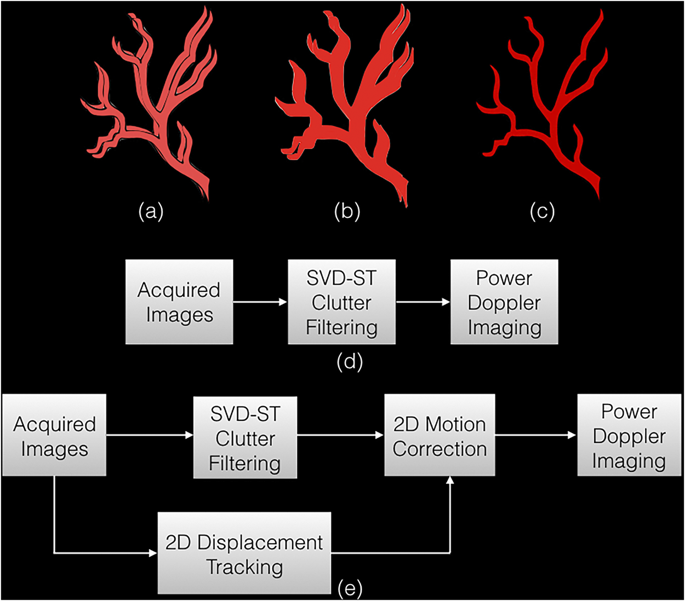
Singular value based spatiotemporal clutter filtering (SVD-STF) can significantly improve the sensitivity of blood flow imaging in small vessels without using contrast agents. However, despite effective clutter filtering, large physiological motion in thyroid imaging can impact coherent integration of the Doppler signal and degrade the visualization of the underlying vasculature. In this study, we hypothesize that motion correction of the clutter filtered Doppler ensemble, prior to the power Doppler estimation, can considerably improve the visualization of smalls vessels in suspicious thyroid nodules. We corroborated this hypothesis by conducting in vivo experiments on 10 female patients in the age group 44-82 yrs, with at least one thyroid nodule suspicious of malignancy, with recommendation for fine needle aspiration biopsy. Ultrasound images were acquired using a clinical ultrasound scanner, implemented with compounded plane wave imaging. Axial and lateral displacements associated with the thyroid nodules were estimated using 2D normalized cross-correlation. Subsequently, the tissue clutter associated with the Doppler ensemble was suppressed using SVD-STF. Motion correction of the clutter-filtered Doppler ensemble was achieved using a spline based sub-pixel interpolation. The results demonstrated that power Doppler images of thyroid nodules were noticeably degraded due to large physiological motion of the pulsating carotid artery in the proximity. The resultant power Doppler images were corrupted with signal distortion, motion blurring and occurrence of artificial shadow vessels and displayed visibly low signal-to-background contrast. In contrast, the power Doppler images obtained from the motion corrected ultrasound data addressed the issue and considerabley improved the visualization of blood flow. The signal-to-noise ratio and the contrast-to-noise ratio increased by up to 15.2 dB and 12.1 dB, respectively. Across the ten subjects, the highest improvement was observed for the nodule with the largest motion. These preliminary results show the ability of using motion correction to improve the visualization of small vessel blood flow in thyroid, without using any contrast agents. The results of this feasibility study were encouraging, and warrant further development and more in vivo validation in moving tissues and organs.
中文翻译:

使用运动校正功率多普勒成像的基于非造影剂的甲状腺小血管成像。
基于奇异值的时空杂波滤波(SVD-STF)可以显着提高小血管中血流成像的灵敏度,而无需使用造影剂。然而,尽管进行了有效的杂波过滤,甲状腺成像中的较大生理运动仍会影响多普勒信号的相干积分并降低基础脉管系统的可视性。在这项研究中,我们假设在功率多普勒估计之前对杂波滤波后的多普勒集合进行运动校正可以显着改善可疑甲状腺结节中小血管的可视化。我们通过对年龄在44-82岁的10名女性患者进行体内实验,证实了这一假设,其中至少一个甲状腺结节可疑为恶性,建议进行细针穿刺活检。使用临床超声扫描仪获取超声图像,并通过复合平面波成像实现。使用2D归一化互相关估计与甲状腺结节相关的轴向和横向位移。随后,使用SVD-STF抑制了与多普勒合奏相关的组织杂波。使用基于样条的子像素插值可实现对杂波滤波后的多普勒合奏的运动校正。结果表明,由于附近的搏动性颈动脉的较大生理运动,甲状腺结节的功率多普勒图像明显降低。产生的功率多普勒图像因信号失真,运动模糊和人造阴影血管的出现而损坏,并显示出明显的信噪比。相比之下,从运动校正后的超声数据获得的功率多普勒图像解决了该问题,并极大地改善了血流的可视性。信噪比和对比度噪声比分别增加了15.2 dB和12.1 dB。在十个受试者中,最大运动的结节观察到了最大的改善。这些初步结果表明,使用运动校正可以改善甲状腺中小血管血流的可视化,而无需使用任何造影剂。这项可行性研究的结果令人鼓舞,并需要进一步发展,并需要在运动的组织和器官中进行更多的体内验证。信噪比和对比度噪声比分别增加了15.2 dB和12.1 dB。在十个受试者中,最大运动的结节观察到了最大的改善。这些初步结果表明,使用运动校正可以改善甲状腺中小血管血流的可视化,而无需使用任何造影剂。这项可行性研究的结果令人鼓舞,并需要进一步发展,并需要在运动的组织和器官中进行更多的体内验证。信噪比和对比度噪声比分别增加了15.2 dB和12.1 dB。在十个受试者中,最大运动的结节观察到了最大的改善。这些初步结果表明,使用运动校正可以改善甲状腺中小血管血流的可视化,而无需使用任何造影剂。这项可行性研究的结果令人鼓舞,并需要进一步发展,并需要在运动的组织和器官中进行更多的体内验证。无需使用任何造影剂。这项可行性研究的结果令人鼓舞,并需要进一步发展,并需要在运动的组织和器官中进行更多的体内验证。不使用任何造影剂。这项可行性研究的结果令人鼓舞,并需要进一步发展,并需要在运动的组织和器官中进行更多的体内验证。
更新日期:2018-10-17
中文翻译:
使用运动校正功率多普勒成像的基于非造影剂的甲状腺小血管成像。
基于奇异值的时空杂波滤波(SVD-STF)可以显着提高小血管中血流成像的灵敏度,而无需使用造影剂。然而,尽管进行了有效的杂波过滤,甲状腺成像中的较大生理运动仍会影响多普勒信号的相干积分并降低基础脉管系统的可视性。在这项研究中,我们假设在功率多普勒估计之前对杂波滤波后的多普勒集合进行运动校正可以显着改善可疑甲状腺结节中小血管的可视化。我们通过对年龄在44-82岁的10名女性患者进行体内实验,证实了这一假设,其中至少一个甲状腺结节可疑为恶性,建议进行细针穿刺活检。使用临床超声扫描仪获取超声图像,并通过复合平面波成像实现。使用2D归一化互相关估计与甲状腺结节相关的轴向和横向位移。随后,使用SVD-STF抑制了与多普勒合奏相关的组织杂波。使用基于样条的子像素插值可实现对杂波滤波后的多普勒合奏的运动校正。结果表明,由于附近的搏动性颈动脉的较大生理运动,甲状腺结节的功率多普勒图像明显降低。产生的功率多普勒图像因信号失真,运动模糊和人造阴影血管的出现而损坏,并显示出明显的信噪比。相比之下,从运动校正后的超声数据获得的功率多普勒图像解决了该问题,并极大地改善了血流的可视性。信噪比和对比度噪声比分别增加了15.2 dB和12.1 dB。在十个受试者中,最大运动的结节观察到了最大的改善。这些初步结果表明,使用运动校正可以改善甲状腺中小血管血流的可视化,而无需使用任何造影剂。这项可行性研究的结果令人鼓舞,并需要进一步发展,并需要在运动的组织和器官中进行更多的体内验证。信噪比和对比度噪声比分别增加了15.2 dB和12.1 dB。在十个受试者中,最大运动的结节观察到了最大的改善。这些初步结果表明,使用运动校正可以改善甲状腺中小血管血流的可视化,而无需使用任何造影剂。这项可行性研究的结果令人鼓舞,并需要进一步发展,并需要在运动的组织和器官中进行更多的体内验证。信噪比和对比度噪声比分别增加了15.2 dB和12.1 dB。在十个受试者中,最大运动的结节观察到了最大的改善。这些初步结果表明,使用运动校正可以改善甲状腺中小血管血流的可视化,而无需使用任何造影剂。这项可行性研究的结果令人鼓舞,并需要进一步发展,并需要在运动的组织和器官中进行更多的体内验证。无需使用任何造影剂。这项可行性研究的结果令人鼓舞,并需要进一步发展,并需要在运动的组织和器官中进行更多的体内验证。不使用任何造影剂。这项可行性研究的结果令人鼓舞,并需要进一步发展,并需要在运动的组织和器官中进行更多的体内验证。


 京公网安备 11010802027423号