Our official English website, www.x-mol.net, welcomes your
feedback! (Note: you will need to create a separate account there.)
Quantitative vessel tortuosity: A potential CT imaging biomarker for distinguishing lung granulomas from adenocarcinomas.
Scientific Reports ( IF 3.8 ) Pub Date : 2018-Oct-16 , DOI: 10.1038/s41598-018-33473-0 Mehdi Alilou 1 , Mahdi Orooji 1 , Niha Beig 1 , Prateek Prasanna 1 , Prabhakar Rajiah 2 , Christopher Donatelli 2 , Vamsidhar Velcheti 3 , Sagar Rakshit 3 , Michael Yang 2 , Frank Jacono 4 , Robert Gilkeson 2 , Philip Linden 2 , Anant Madabhushi 1
Scientific Reports ( IF 3.8 ) Pub Date : 2018-Oct-16 , DOI: 10.1038/s41598-018-33473-0 Mehdi Alilou 1 , Mahdi Orooji 1 , Niha Beig 1 , Prateek Prasanna 1 , Prabhakar Rajiah 2 , Christopher Donatelli 2 , Vamsidhar Velcheti 3 , Sagar Rakshit 3 , Michael Yang 2 , Frank Jacono 4 , Robert Gilkeson 2 , Philip Linden 2 , Anant Madabhushi 1
Affiliation
|
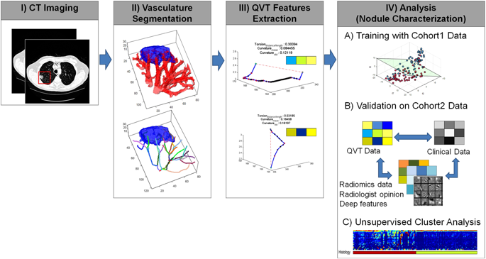
Adenocarcinomas and active granulomas can both have a spiculated appearance on computed tomography (CT) and both are often fluorodeoxyglucose (FDG) avid on positron emission tomography (PET) scan, making them difficult to distinguish. Consequently, patients with benign granulomas are often subjected to invasive surgical biopsies or resections. In this study, quantitative vessel tortuosity (QVT), a novel CT imaging biomarker to distinguish between benign granulomas and adenocarcinomas on routine non-contrast lung CT scans is introduced. Our study comprised of CT scans of 290 patients from two different institutions, one cohort for training (N = 145) and the other (N = 145) for independent validation. In conjunction with a machine learning classifier, the top informative and stable QVT features yielded an area under receiver operating characteristic curve (ROC AUC) of 0.85 in the independent validation set. On the same cohort, the corresponding AUCs for two human experts including a radiologist and a pulmonologist were found to be 0.61 and 0.60, respectively. QVT features also outperformed well known shape and textural radiomic features which had a maximum AUC of 0.73 (p-value = 0.002), as well as features learned using a convolutional neural network AUC = 0.76 (p-value = 0.028). Our results suggest that QVT features could potentially serve as a non-invasive imaging biomarker to distinguish granulomas from adenocarcinomas on non-contrast CT scans.
中文翻译:

定量血管迂曲度:一种潜在的 CT 成像生物标志物,用于区分肺肉芽肿和腺癌。
腺癌和活动性肉芽肿在计算机断层扫描 (CT) 上都可能具有毛刺状外观,并且在正电子发射断层扫描 (PET) 扫描中通常都对氟脱氧葡萄糖 (FDG) 感兴趣,因此难以区分。因此,良性肉芽肿患者经常接受侵入性手术活检或切除术。在这项研究中,定量血管迂曲度 (QVT) 是一种新型 CT 成像生物标志物,可在常规非对比肺 CT 扫描中区分良性肉芽肿和腺癌。我们的研究包括来自两个不同机构的 290 名患者的 CT 扫描,一组用于培训 (N = 145),另一组 (N = 145) 用于独立验证。结合机器学习分类器,在独立验证集中,信息量最大且稳定的 QVT 特征产生的受试者工作特征曲线下面积 (ROC AUC) 为 0.85。在同一队列中,包括放射科医师和肺科医师在内的两位人类专家的相应 AUC 分别为 0.61 和 0.60。QVT 特征也优于众所周知的形状和纹理放射组学特征,其最大 AUC 为 0.73(p 值 = 0.002),以及使用卷积神经网络学习的特征 AUC = 0.76(p 值 = 0.028)。我们的结果表明,QVT 特征可能作为一种非侵入性成像生物标志物,在非对比 CT 扫描中区分肉芽肿和腺癌。两个人类专家(包括放射科医师和肺科医师)的相应 AUC 分别为 0.61 和 0.60。QVT 特征也优于众所周知的形状和纹理放射组学特征,其最大 AUC 为 0.73(p 值 = 0.002),以及使用卷积神经网络学习的特征 AUC = 0.76(p 值 = 0.028)。我们的结果表明,QVT 特征可能作为一种非侵入性成像生物标志物,在非对比 CT 扫描中区分肉芽肿和腺癌。两个人类专家(包括放射科医师和肺科医师)的相应 AUC 分别为 0.61 和 0.60。QVT 特征也优于众所周知的形状和纹理放射组学特征,其最大 AUC 为 0.73(p 值 = 0.002),以及使用卷积神经网络学习的特征 AUC = 0.76(p 值 = 0.028)。我们的结果表明,QVT 特征可能作为一种非侵入性成像生物标志物,在非对比 CT 扫描中区分肉芽肿和腺癌。
更新日期:2018-10-16
中文翻译:
定量血管迂曲度:一种潜在的 CT 成像生物标志物,用于区分肺肉芽肿和腺癌。
腺癌和活动性肉芽肿在计算机断层扫描 (CT) 上都可能具有毛刺状外观,并且在正电子发射断层扫描 (PET) 扫描中通常都对氟脱氧葡萄糖 (FDG) 感兴趣,因此难以区分。因此,良性肉芽肿患者经常接受侵入性手术活检或切除术。在这项研究中,定量血管迂曲度 (QVT) 是一种新型 CT 成像生物标志物,可在常规非对比肺 CT 扫描中区分良性肉芽肿和腺癌。我们的研究包括来自两个不同机构的 290 名患者的 CT 扫描,一组用于培训 (N = 145),另一组 (N = 145) 用于独立验证。结合机器学习分类器,在独立验证集中,信息量最大且稳定的 QVT 特征产生的受试者工作特征曲线下面积 (ROC AUC) 为 0.85。在同一队列中,包括放射科医师和肺科医师在内的两位人类专家的相应 AUC 分别为 0.61 和 0.60。QVT 特征也优于众所周知的形状和纹理放射组学特征,其最大 AUC 为 0.73(p 值 = 0.002),以及使用卷积神经网络学习的特征 AUC = 0.76(p 值 = 0.028)。我们的结果表明,QVT 特征可能作为一种非侵入性成像生物标志物,在非对比 CT 扫描中区分肉芽肿和腺癌。两个人类专家(包括放射科医师和肺科医师)的相应 AUC 分别为 0.61 和 0.60。QVT 特征也优于众所周知的形状和纹理放射组学特征,其最大 AUC 为 0.73(p 值 = 0.002),以及使用卷积神经网络学习的特征 AUC = 0.76(p 值 = 0.028)。我们的结果表明,QVT 特征可能作为一种非侵入性成像生物标志物,在非对比 CT 扫描中区分肉芽肿和腺癌。两个人类专家(包括放射科医师和肺科医师)的相应 AUC 分别为 0.61 和 0.60。QVT 特征也优于众所周知的形状和纹理放射组学特征,其最大 AUC 为 0.73(p 值 = 0.002),以及使用卷积神经网络学习的特征 AUC = 0.76(p 值 = 0.028)。我们的结果表明,QVT 特征可能作为一种非侵入性成像生物标志物,在非对比 CT 扫描中区分肉芽肿和腺癌。


 京公网安备 11010802027423号