Gastrointestinal Endoscopy ( IF 6.7 ) Pub Date : 2018-10-13 , DOI: 10.1016/j.gie.2018.09.037 Takahide Shinagawa , Keisuke Hata , Teppei Morikawa , Hirotoshi Takiyama , Shigenobu Emoto , Koji Murono , Manabu Kaneko , Kazuhito Sasaki , Takeshi Nishikawa , Toshiaki Tanaka , Kazushige Kawai , Masashi Fukayama , Hiroaki Nozawa
|
Background and Aims
The appropriate site for targeted biopsy during surveillance colonoscopy for ulcerative colitis (UC) is still unclear. We aimed to clarify key endoscopic findings suggestive of neoplastic lesions for targeted biopsy in UC.
Methods
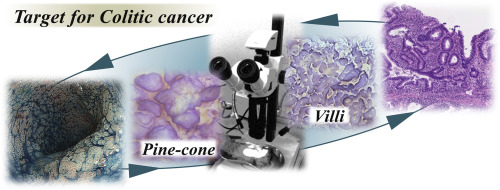
First, we created 769 stereomicroscopic pictures (509 neoplastic, 260 non-neoplastic) mimicking magnifying colonoscopic images from surgically resected specimens, including areas surrounding 25 neoplastic lesions in 15 patients with colitis-associated cancer at a single referral center. Second, we validated the results by using 113 magnifying endoscopic images (64 neoplastic, 49 non-neoplastic) from 39 lesions in 26 patients. Two evaluators, blinded to the pathologic diagnosis, independently classified them according to Kudo’s pit pattern and surface morphology, such as pine-cone/villi patterns. The correlation between stereomicroscopic and pathologic findings (neoplastic vs non-neoplastic) for each image was investigated. The interobserver agreement was assessed using kappa statistics.
Results
In the stereomicroscopic analysis, neoplastic pit patterns (types III-V) were significantly correlated with the presence of neoplasia (sensitivity 77.4%, specificity 89.5%, kappa value 0.677). Pine-cone/villi patterns also showed high specificity (96.8%) but low sensitivity (21.4%, kappa value 0.625) for neoplasia. Endoscopic validation showed similar trends. A revision of the endoscopic findings of flat dysplasia with non-neoplastic pit patterns revealed that a reddish area may facilitate the identification of such lesions.
Conclusions
Targeted biopsies are recommended, especially for lesions showing pine-cone/villi patterns in addition to neoplastic pit patterns. For flat “non-neoplastic pit patterns,” a reddish area may be an indication for a biopsy.
中文翻译:
松锥状和绒毛状是内窥镜征象,提示溃疡性结肠炎相关的大肠癌和不典型增生
背景和目标
溃疡性结肠炎(UC)监测结肠镜检查期间进行靶向活检的合适部位仍不清楚。我们旨在阐明关键的内窥镜检查结果,提示在UC靶向活检中有肿瘤性病变。
方法
首先,我们创建了769幅立体显微镜图片(509例肿瘤肿瘤,260例非肿瘤肿瘤),模拟了从手术切除标本中放大的结肠镜图像,包括在单个转诊中心的15例结肠炎相关癌症患者的25例肿瘤病变区域。第二,我们使用来自26例患者的39个病变的113幅放大内窥镜图像(64例肿瘤,49例非肿瘤)验证了结果。两名不愿进行病理诊断的评估人员根据工藤的凹坑图案和表面形态(例如松果/绒毛图案)对它们进行了独立分类。研究了每个图像的立体显微镜和病理结果(肿瘤性与非肿瘤性)之间的相关性。观察员之间的协议使用kappa统计数据进行了评估。
结果
在立体显微镜分析中,肿瘤性凹坑模式(III-V型)与肿瘤的存在显着相关(敏感性为77.4%,特异性为89.5%,κ值为0.677)。松果/绒毛模式对瘤形成也显示出高特异性(96.8%),但敏感性低(21.4%,κ值0.625)。内镜验证显示出相似的趋势。对具有非肿瘤性凹坑型的扁平不典型增生的内窥镜检查结果进行的修订显示,红色区域可能有助于识别此类病变。
结论
建议进行有针对性的活检,尤其是对于有松果/绒毛样病变的病变,以及肿瘤性凹坑型病变。对于平坦的“非肿瘤性凹坑型”,红色区域可能是活检的指征。


 京公网安备 11010802027423号