Our official English website, www.x-mol.net, welcomes your feedback! (Note: you will need to create a separate account there.)
Bio‐Inspired Micropatterned Platforms Recapitulate 3D Physiological Morphologies of Bone and Dentinal Cells
Advanced Science ( IF 15.1 ) Pub Date : 2018-10-12 , DOI: 10.1002/advs.201801037 Chi Ma 1 , Bei Chang 1 , Yan Jing 2 , Harry Kim 3 , Xiaohua Liu 1
Advanced Science ( IF 15.1 ) Pub Date : 2018-10-12 , DOI: 10.1002/advs.201801037 Chi Ma 1 , Bei Chang 1 , Yan Jing 2 , Harry Kim 3 , Xiaohua Liu 1
Affiliation

|
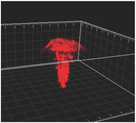
Cells exhibit distinct 3D morphologies in vivo, and recapitulation of physiological cell morphologies in vitro is pivotal not only to elucidate many fundamental biological questions, but also to develop new approaches for tissue regeneration and drug screening. However, conventional cell culture methods in either a 2D petri dish or a 3D scaffold often lead to the loss of the physiological morphologies for many cells, such as bone cells (osteocytes) and dentinal cells (odontoblasts). Herein, a unique approach in developing a 3D extracellular matrix (ECM)‐like micropatterned synthetic matrix as a physiologically relevant 3D platform is reported to recapitulate the morphologies of osteocytes and odontoblasts in vitro. The bio‐inspired micropatterned matrix precisely mimics the hierarchic 3D nanofibrous tubular/canaliculi architecture as well as the compositions of the ECM of mineralized tissues, and is capable of controlling one single cell in a microisland of the matrix. Using this bio‐inspired 3D platform, individual bone and dental stem cells are successfully manipulated to recapitulate the physiological morphologies of osteocytes and odontoblasts in vitro, respectively. This work provides an excellent platform for an in‐depth understanding of cell–matrix interactions in 3D environments, paving the way for designing next‐generation biomaterials for tissue regeneration.
中文翻译:

仿生微图案平台重现骨和牙本质细胞的 3D 生理形态
细胞在体内表现出独特的 3D 形态,在体外重现生理细胞形态不仅对于阐明许多基本生物学问题至关重要,而且对于开发组织再生和药物筛选的新方法也至关重要。然而,2D 培养皿或 3D 支架中的传统细胞培养方法通常会导致许多细胞生理形态的丧失,例如骨细胞(骨细胞)和牙本质细胞(成牙本质细胞)。据报道,开发 3D 细胞外基质 (ECM) 类微图案合成基质作为生理相关 3D 平台的独特方法可在体外重现骨细胞和成牙本质细胞的形态。这种仿生微图案基质精确模拟了分层的 3D 纳米纤维管状/小管结构以及矿化组织 ECM 的成分,并且能够控制基质微岛中的单个细胞。利用这种仿生 3D 平台,可以成功地操纵单个骨干细胞和牙齿干细胞,分别在体外重现骨细胞和成牙本质细胞的生理形态。这项工作为深入了解 3D 环境中的细胞-基质相互作用提供了一个极好的平台,为设计下一代组织再生生物材料铺平了道路。
更新日期:2018-10-12
中文翻译:

仿生微图案平台重现骨和牙本质细胞的 3D 生理形态
细胞在体内表现出独特的 3D 形态,在体外重现生理细胞形态不仅对于阐明许多基本生物学问题至关重要,而且对于开发组织再生和药物筛选的新方法也至关重要。然而,2D 培养皿或 3D 支架中的传统细胞培养方法通常会导致许多细胞生理形态的丧失,例如骨细胞(骨细胞)和牙本质细胞(成牙本质细胞)。据报道,开发 3D 细胞外基质 (ECM) 类微图案合成基质作为生理相关 3D 平台的独特方法可在体外重现骨细胞和成牙本质细胞的形态。这种仿生微图案基质精确模拟了分层的 3D 纳米纤维管状/小管结构以及矿化组织 ECM 的成分,并且能够控制基质微岛中的单个细胞。利用这种仿生 3D 平台,可以成功地操纵单个骨干细胞和牙齿干细胞,分别在体外重现骨细胞和成牙本质细胞的生理形态。这项工作为深入了解 3D 环境中的细胞-基质相互作用提供了一个极好的平台,为设计下一代组织再生生物材料铺平了道路。


 京公网安备 11010802027423号